Download
1 / 1
30 likes | 204 Vues
Sulforaphane modulates antioxidant redox signalling in human aortic adventitial fibroblasts Tabasum Mughal 1 , Jonathan Wong 1 , Giovanni E Mann 1 , Maddy Parsons 2 & Richard CM Siow 1

E N D
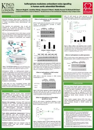
Sulforaphane modulates antioxidant redox signalling in human aortic adventitial fibroblasts Tabasum Mughal1, Jonathan Wong1, Giovanni E Mann1, Maddy Parsons2 & Richard CM Siow1 1Cardiovascular Division, School of Medicine, 2 Randall Division of Cell and Molecular Biophysics, King’s College London INTRODUCTION RESULTS SFN (10 mM) causes an initial decrease in total glutathione levels in HAoAF with a significant decrease at 4 h. Total GSH levels return to basal levels by 24 h. Adventitial fibroblast differentiation, proliferation and migration is a characteristic event and plays a key role in vessel remodelling during atherosclerosis and restenosis1. This migration and proliferation may, in part, be mediated by an enhanced generation of reactive oxygen species (ROS) occurring within of the vessel wall during disease progression2. Nrf2 is a key transcription factor which plays a central role in the induction of many cytoprotective genes such as glutathione (GSH), heme oxygenase-1 (HO-1) and (NQO-1) in response to electrophillic and oxidative stress3. Sulforaphane (SFN), an isothiocyanate present in cruciferous vegetables such as broccoli, protects cells against oxidative stress and inflammation through the activation of Nrf24. Dietary SFN may therefore confer protection against ROS-mediated vascular cell dysfunction and remodelling in atherosclerosis through attenuating phenotypic modulation of adventitial fibroblast to myofibroblasts. In this study we have investigated whether SFN modulates redox signalling (HO-1, NQO-1 and GSH levels) in human aortic adventitial fibroblasts. Effect of sulforaphane on HO-1 and NQO1 expression A SFN (10 µM) elicits the induction of HO-1 at 8 h and causes a dose-dependent increase in NQO1 expression at 8 and 24 h. A * Control SFN A Time (h) 4 8 24 4 8 24 B HO-1 B Tubulin B * Control * SFN (10 µM) Figure 5. Effect of SFN on total glutathione levels in HAoAF. Cells were treated with SFN (10 µM, 0-24 h) and levels of glutathione were determined using a fluorescence assay and expressed relative to cellular protein content. (A) Total intracellular glutathione levels in HAoAF (B) Percentage change in GSH levels compared to untreated control. n= 3 independent cell cultures. *p<0.05 (Sulforaphane) 4 8 24 Time (h) C 1 µM 2.5 µM 10 µM Control Vehicle 5 µM . Effect of sulforaphane on pAkt expression Time (h) 8 24 8 24 8 24 8 24 8 24 8 24 SFN (10 mM) causes a time dependent increase in Aktphosphorylation, with a marked increased by 4 h. NQO1 (HO-1) Tubulin Control SFN (10 µM) Figure 2.Antioxidant protein expression in HAoAF treated with SFN (1 - 10 µM). Cells were treated with SFN (1 - 10 µM) for 4, 8 or 24 h (A) HO-1 (32 kDa) and (C) NQO1 (31 kDa) expression was determined by western blot analysis with α-tubulin (55 kDa) used as a protein loading control. (B) Relative band densities expressed as a ratio of HO-1/α-tubulin. Immunoblots shown are representative of data from n=3 independent cell cultures. *p<0.05 Time (h) 0.5 1 2 4 0.5 1 2 4 pAkt Figure 1. The Nrf2-Keap1 pathway. Nrf2 is a key transcription factor involved in the induction of key cytoprotective genes in response to oxidative or electrophillic stress. Adapted from Taguchi et al., 2011. Tubulin Effect of sulforaphane on Nrf2 nuclear translocation • Fig 6. pAkt protein expression in HAoAF treated with SFN (10 µM) • Cells were treated with SFN (10 µM, 30 min - 4 h). pAkt (60 kDa) • expression was determined by western blot analysis with α-tubulin (55 • kDa) used as a protein loading control. Immunoblots shown are • representative of data from n=3 independent cell cultures SFN elicits a two-fold increase in Nrf2 nuclear translocation at 2 h. Cytoplasmic Nuclear Ctrl TGF-B SFN Ctrl TGF-B SFN CONCLUSIONS Nrf2 110 kDa Lamin A SFN may protect HAoAF against oxidative stress in via activation of the Nrf2 pathway to elicit an increase in HO-1 and NQO1 expression. SFN also causes a time-dependent increase in Aktphosphorylation, suggesting that it may be upstream of the Nrf2 pathway It is possible that the acute decrease in total glutathione levels drives induction of the Nrf2 pathway. These results suggest dietary SFN may reduce vascular remodelling through a reduction in oxidative stress mediated changes in HAoAF viability and phenotype. Tubulin Figure 3.Nrf2 nuclear expression in HAoAF treated with SFN. Cells were treated with SFN (10 µM) for 2 h and Nrf2 nuclear translocation was determined by western blot analysis with Lamin A (70 kDa) and α-tubulin (55 kDa) used as protein loading controls for nuclear and cytoplasmic fractions respectively. METHODS Cell culture: Primary human aortic adventitial fibroblasts (HAoAF, at passage 4-9) were cultured in phenol red-free DMEM containing 10% fetal calf serum (FCS) and equilibrated overnight in DMEM (1% FCS) prior to treatments (0-24 h) with SFN (1-10 µM). Western blot analyses: Following SFN treatments expression of HO-1, NQO1 and Nrf2 was determined in whole cell lysates and nuclear extracts by immunoblot analyses using specific primary antibodies and HRP-conjugated secondary antibodies. Expression of tubulin and lamin A/C were determined to provide an index of protein loading. Total cellular glutathione measurement: Following SFN treatments cells were incubated with tri-chloroacetic acid (6.5%) for 10 minutes. Glutathione levels in the supernatants were determined using the orthophthaldehyde fluorescence assay and were expressed relative to cellular protein content. Statistical analysis: Data was analysed using the Students paired t-test with p<0.05 considered statistically significant. C Effect of sulforaphane on intracellular glutathione levels SFN (5 mM) causes a significant depletion in total glutathione levels in HAoAF at 2 and 4 h. GSH levels return to basal levels by 24 h. REFERENCES ** Mailero and Taylor (2007) The role of the adventitia in vascular inflammation. Cardiovasc Res 75: 640-648. Haurani and Pagano (2007) Adventital fibroblast reactive oxygen species as autocrine and paracrine mediators of remodelling: bellweather for vascular disease? Cardiovasc Res 75: 679-689 Taguchi et al. (2011) Molecular mechanisms of the Keap1-Nrf2 pathway in stress response and cancer evolution Genes Cells 16: 123-40 Cheung and Kong (2010) Molecular targets of dietary phenethylisothiocyanate and sulforaphane for cancer chemoprevention AAPS1: 87-97 This research was supported by the British Heart Foundation Figure 4.Glutathione levels in HAoAF treated with SFN (5 µM). Cells were treated with SFN (5 µM) for 2, 4, 8, 24 and 36 h and subsequently incubated with 6.5% TCA. Following incubation the supernantant was removed from cells and total glutathione levels were determined using the opthalaldehyde assay. Glutathione was expressed relative to cellular protein content. **p<0.01