Download

1 / 26
270 likes | 854 Vues
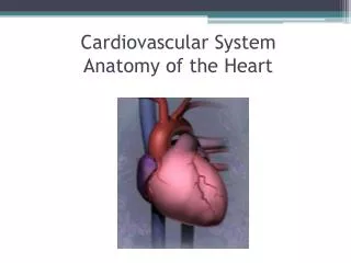
Conductive System Of Heart. Dr. Mahvash Khan MBBS, MPhiL Assistant Professor Physiology Rawal Institute of Health Sciences. The heart has a special system for generating rhythmical electrical impulses to cause rhythmical contraction of the heart muscle.

E N D
Conductive System Of Heart Dr. Mahvash Khan MBBS, MPhiL Assistant Professor Physiology Rawal Institute of Health Sciences
The heart has a special system for • generating rhythmical electrical impulses to cause rhythmical contraction of the heart muscle. • conducting these impulses rapidly through the heart.
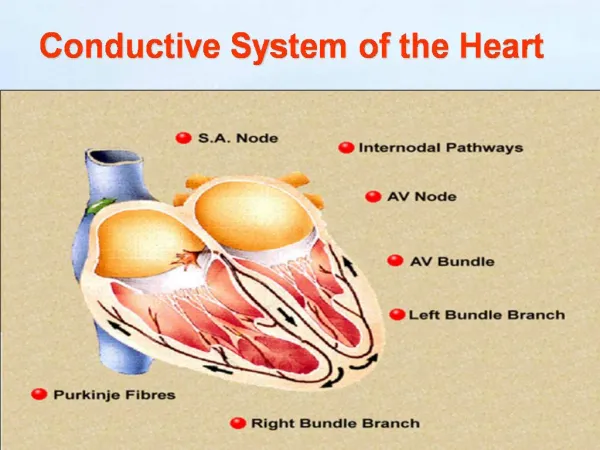
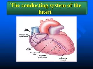
Components Of The Conductive System • SA node • Internodal pathways • A-V node • A-V bundle • Right and Left bundle branches
Automaticity/Rhythmicity • Automaticity means the ability of the cell to undergo depolarization spontaneously causing the production of electrical impulses. • Rhythmicity means that spontaneous depolarization occurs at regular intervals i.e in a rhythmic manner.
Cardiac Impulse • The action potential in the heart is also called the cardiac impulse and like action potential in the nerve fibers it travels.
SA node • SA node is the pacemaker of the heart. Normally SA node is responsible for generating the electrical impulses that bring about the mechanical activity i.e contraction of the heart. • SA node has the fastest rate of autorhythmicity.
Location of the SA node • SA node is a small, ellipsoid strip of specialized cardiac muscle about 3mm wide, 15 mm long, and 1mm thick. It is located in the superior posterolateral wall of the right atrium immediately below and slightly lateral to the opening of the superior venacava. • SA nodal fibers connect directly with the atrial muscle fibers.
Spread of Cardiac Impulse from SA node to Atrial muscle The cardiac impulse after it’s origin in the SA node spreads through out the atrial muscle through two routes • Ordinary Atrial muscle fibers • Specialized anterior, middle and posterior conducting bundles • Anterior internodal bundle of Bachman • Middle internodal bundle of Wenkebach • Posterior internodal bundle of Thoral
These inter nodal pathways conduct the impulses at a faster rate than the ordinary atrial muscle fibers. • The cause of rapid conduction in these bundles is the presence of specialized conduction fibers.
The velocity of conduction in most atrial muscle is about 0.3m/sec. • In the specialized internodal pathways the conduction velocity may reach upto 1m/sec. • The impulse after leaving SA node takes 0.03 sec to reach the AV node.
AV node The AV node is located in the posterior wall of the right atrium immediately behind the tricuspid valve.
AV nodal Delay • Due to the slow conduction in AV node there is a delay of 0.09 sec in the AV node, before the impulse reaches the penetrating portion of AV bundle. • There is a delay of 0.04 sec in the penetrating portion of AV bundle. • There is a total delay of 0.13 sec in the AV node and AV bundle system. • There is a delay of 0.03 sec from the sinus node to the AV node, thus making total delay of 0.16 sec before the excitatory signal finally reaches the contracting muscle of ventricles.
Cause of Slow Conduction in the A-V Node The cause of slow conduction is mainly diminished number of gap junctions between the successive cells in the conducting pathways. As a result of which there is great resistance to conduction of excitatory ions from one conducting fiber to the next.
Significance of AV nodal delay • The cardiac impulse does not travel from the atria to the ventricles too rapidly. • This delay allows time for the atria to empty their blood into the ventricles before ventricular contraction begins. This increases the efficiency of the pumping action of the heart. • It is primarily the AV node and it’s adjacent fibers that delay this transmission into the ventricles
AV Bundle or Bundle of His • From the AV node arises a special conducting pathway called the bundle of His. Except for the very small part which penetrates through the AV fibrous tissue and has low conduction velocity, the bundle of His is made up of purkinje fibers which possess maximum conduction velocity in the heart.
Purkinje fibers are very large fibers and they transmit action potentials at a velocity of 1.5 to 4.0 m/sec. • The rapid transmission of action potentials through the Purkinje fibers is believed to be caused by a very high level of permeability of gap junctions at the intercalated discs between the successive cells of Purkinje fibers. • The rapid conduction through the purkinje fibers ensures that different parts of ventricles are excited almost simultaneously; this greatly increases the efficiency of heart as a pump.
Normally the Bundle of His is the only conducting mass between the atrial and ventricular musculature and it transmits the cardiac impulses from the AV node to the ventricles.
Right and Left Bundle Branches • After penetrating the fibrous tissue between the atrial and ventricular muscle, the distal portion of the A-V bundle passes downward in the ventricular septum for 5 to 15 mm toward the apex of the heart. • Then the bundle of His splits into two branches which are called right and left bundle branches that lie on the respective sides of the ventricular septum.
Each branch spreads downward toward the apex of the ventricle, progressively dividing into smaller branches. • These branches inturn course sidewise around each ventricular chamber and back toward the base of heart. • The ends of Purkinje fibers penetrate about one third of the way into muscle mass and finally become continuous with cardiac muscle fibers.
From the time the cardiac impulse enters the bundle branches until it reaches the terminations of Purkinje fibers , the total elapsed time averages only 0.03 sec.
One- way Conduction through AV bundle • A special characteristic of the A-V bundle is it’s inability, except in the abnormal states , of action potentials to travel backward from the ventricles to the atria. • This prevents re-entry of cardiac impulse by this route from the ventricles to the atria. • The atrial muscle is separated from the ventricular muscle by a continuous fibrous barrier which acts as an insulator to prevent the passage of cardiac impulse between the atrial and ventricular muscle through any other route besides forward conduction through A-V bundle itself.
Conduction in the Cardiac Muscle • Once the impulse reaches the ends of the Purkinje fibers it is transmitted through the ventricular muscle mass by the ventricular muscle fibers themselves. • For transmission of the cardiac impulse from the endocardial surface to the epicardial surface recquires another 0.03 sec. • Thus the total time for transmission of cardiac impulse from the initial bundle branches to the last of the ventricular muscle fibers in the normal heart is about 0.06 sec.
Normal Rate of Action potential Discharge in Autorhythmic Tissues of the Heart