Download
1 / 27
300 likes | 639 Vues
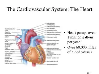
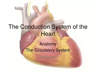
The Electrical System of the Heart. Cardiac Muscle Contraction. Depolarization of the heart is rhythmic and spontaneous About 1% of cardiac cells have automaticity— (are self-excitable) Gap junctions ensure the heart contracts as a unit Long absolute refractory period (250 ms). 1. Action

E N D
Cardiac Muscle Contraction • Depolarization of the heart is rhythmic and spontaneous • About 1% of cardiac cells have automaticity— (are self-excitable) • Gap junctions ensure the heart contracts as a unit • Long absolute refractory period (250 ms)
1 Action potential Depolarizationis due to Na+influx through fast voltage-gated Na+ channels. A positive feedback cycle rapidly opens many Na+ channels, reversing the membrane potential. Channel inactivation ends this phase. Plateau 2 Tension development (contraction) Membrane potential (mV) 3 Tension (g) 1 2 Plateau phaseis due to Ca2+ influx through slow Ca2+channels. This keeps the cell depolarized because few K+channels are open. Absolute refractory period 3 Repolarizationis due to Ca2+channels inactivating and K+ channels opening. This allows K+efflux, which brings the membrane potential back to its resting voltage. Time (ms) Figure 18.12
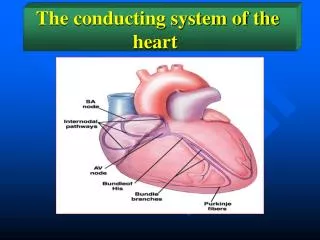
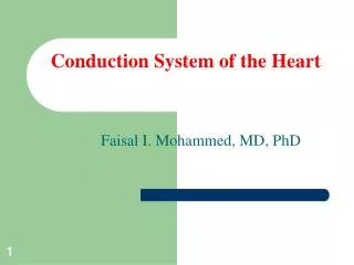
Heart Physiology: Electrical Events Don’t copy • Intrinsic cardiac conduction system • A network of noncontractile (autorhythmic) cells that initiate and distribute impulses to coordinate the depolarization and contraction of the heart
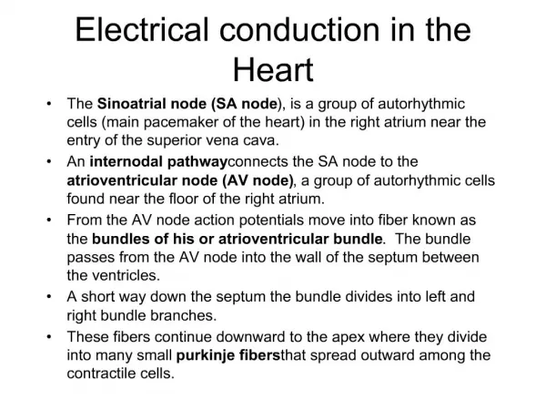
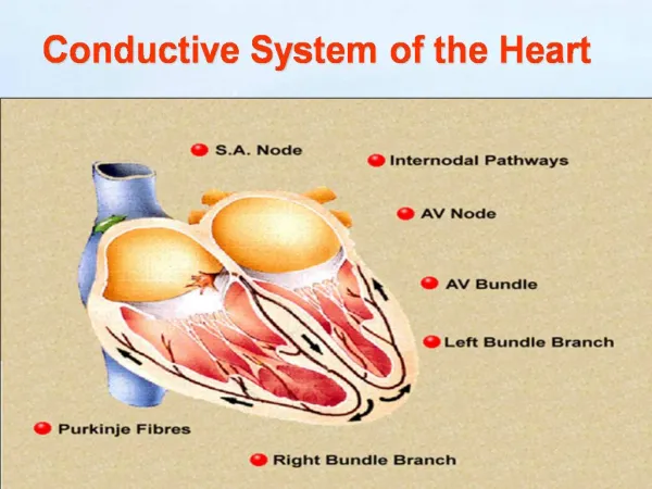
Heart Physiology: Sequence of Excitation • Sinoatrial (SA) node (pacemaker) • Generates impulses about 75 times/minute (sinus rhythm) • Depolarizes faster than any other part of the myocardium
Heart Physiology: Sequence of Excitation • Atrioventricular (AV) node • Smaller diameter fibers; fewer gap junctions • Delays impulses approximately 0.1 second • Depolarizes 50 times per minute in absence of SA node input
Heart Physiology: Sequence of Excitation • Atrioventricular (AV) bundle (bundle of His) • Only electrical connection between the atria and ventricles
Superior vena cava Right atrium Thesinoatrial (SA) node(pacemaker) generates impulses. 1 Internodal pathway Left atrium 2 The impulses pause (0.1 s) at the atrioventricular (AV) node. Purkinje fibers Theatrioventricular (AV) bundle connects the atria to the ventricles. 3 Thebundle branches conduct the impulses through the interventricular septum. 4 Inter- ventricular septum ThePurkinje fibers depolarize the contractile cells of both ventricles. 5 (a) Anatomy of the intrinsic conduction system showing the sequence of electrical excitation Figure 18.14a
Homeostatic Imbalances • Defects in the intrinsic conduction system may result in • Arrhythmias: irregular heart rhythms • Uncoordinated atrial and ventricular contractions • Fibrillation: rapid, irregular contractions; useless for pumping blood
Homeostatic Imbalances • Defective SA node may result in • Ectopic focus: abnormal pacemaker takes over • If AV node takes over, there will be a junctional rhythm (40–60 bpm) • Defective AV node may result in • Partial or total heart block • Few or no impulses from SA node reach the ventricles
Extrinsic Innervation of the Heart • Heartbeat is modified by the ANS • Cardiac centers are located in the medulla oblongata • Cardioacceleratory center innervates SA and AV nodes, heart muscle, and coronary arteries through sympathetic neurons • Cardioinhibitory center inhibits SA and AV nodes through parasympathetic fibers in the vagus nerves
Dorsal motor nucleus of vagus The vagus nerve (parasympathetic) decreases heart rate. Cardioinhibitory center Medulla oblongata Cardio- acceleratory center Sympathetic trunk ganglion Thoracic spinal cord Sympathetic trunk Sympathetic cardiac nervesincrease heart rate and force of contraction. AV node SA node Parasympathetic fibers Sympathetic fibers Interneurons Figure 18.15
Electrocardiography • Electrocardiogram (ECG or EKG): a composite of all the action potentials generated by nodal and contractile cells at a given time • Three waves • P wave: depolarization of SA node • QRS complex: ventricular depolarization • T wave: ventricular repolarization
QRS complex Sinoatrial node Ventricular depolarization Ventricular repolarization Atrial depolarization Atrioventricular node S-T Segment P-Q Interval Q-T Interval Figure 18.16
Depolarization Repolarization SA node R R T P T P Q S 1 Atrial depolarization, initiatedby the SA node, causes theP wave. Q S 4 Ventricular depolarizationis complete. R AV node R T P T P Q S Q 2 With atrial depolarizationcomplete, the impulse isdelayed at the AV node. S 5 Ventricular repolarizationbegins at apex, causing theT wave. R R T P T P Q S Q S 3 Ventricular depolarizationbegins at apex, causing theQRS complex. Atrialrepolarization occurs. 6 Ventricular repolarizationis complete. Figure 18.17
R Depolarization SA node Repolarization T P Q S 1 Atrial depolarization, initiated bythe SA node, causes the P wave. Figure 18.17, step 1
R Depolarization SA node Repolarization T P Q S 1 Atrial depolarization, initiated bythe SA node, causes the P wave. R AV node T P Q S 2 With atrial depolarization complete,the impulse is delayed at the AV node. Figure 18.17, step 2
R Depolarization SA node Repolarization T P Q S 1 Atrial depolarization, initiated bythe SA node, causes the P wave. R AV node T P Q S 2 With atrial depolarization complete,the impulse is delayed at the AV node. R T P Q S 3 Ventricular depolarization beginsat apex, causing the QRS complex.Atrial repolarization occurs. Figure 18.17, step 3
Depolarization Repolarization R T P Q S 4 Ventricular depolarization iscomplete. Figure 18.17, step 4
Depolarization Repolarization R T P Q S 4 Ventricular depolarization iscomplete. R T P Q S 5 Ventricular repolarization beginsat apex, causing the T wave. Figure 18.17, step 5
Depolarization Repolarization R T P Q S 4 Ventricular depolarization iscomplete. R T P Q S 5 Ventricular repolarization beginsat apex, causing the T wave. R T P Q S 6 Ventricular repolarization iscomplete. Figure 18.17, step 6
Depolarization Repolarization SA node R R T P T P Q S 1 Atrial depolarization, initiatedby the SA node, causes theP wave. Q S 4 Ventricular depolarizationis complete. R AV node R T P T P Q S Q 2 With atrial depolarizationcomplete, the impulse isdelayed at the AV node. S 5 Ventricular repolarizationbegins at apex, causing theT wave. R R T P T P Q S Q S 3 Ventricular depolarizationbegins at apex, causing theQRS complex. Atrialrepolarization occurs. 6 Ventricular repolarizationis complete. Figure 18.17
(a) Normal sinus rhythm. (b) Junctional rhythm. The SA node is nonfunctional, P waves are absent, and heart is paced by the AV node at 40 - 60 beats/min. (d) Ventricular fibrillation. These chaotic, grossly irregular ECG deflections are seen in acute heart attack and electrical shock. (c) Second-degree heart block. Some P waves are not conducted through the AV node; hence more P than QRS waves are seen. In this tracing, the ratio of P waves to QRS waves is mostly 2:1. Figure 18.18
Heart Sounds • Two sounds (lub-dup) associated with closing of heart valves • First sound occurs as AV valves close and signifies beginning of systole • Second sound occurs when SL valves close at the beginning of ventricular diastole • Heart murmurs: abnormal heart sounds most often indicative of valve problems
In catheter ablation, catheters are threaded through the blood vessels to the inner heart, and electrodes at the catheter tips transmit energy to destroy a small spot of heart tissue.