Download
1 / 15
180 likes | 548 Vues
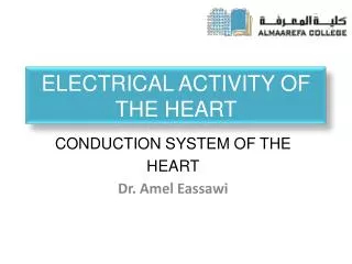
ELECTRICAL ACTIVITY OF THE HEART. Dr. Ayisha Qureshi Assistant Professor MBBS, MPhil. Cardiac cells contract without Nervous Stimulation. . Cardiac muscle, like skeletal muscle & neurons, is an excitable tissue with the ability to generate action potential.

E N D
ELECTRICAL ACTIVITY OF THE HEART Dr. Ayisha Qureshi Assistant Professor MBBS, MPhil.
Cardiac cells contract without Nervous Stimulation. • Cardiac muscle, like skeletal muscle & neurons, is an excitable tissue with the ability to generate action potential. • Most cardiac muscle is contractile (99%), but about 1% of the myocardial cells are specialized to generate action potentials spontaneously. These cells are responsible for a unique property of the heart: its ability to contract without any outside signal. • The heart can contract without an outside signal because the signal for contraction is myogenic, originating within the heart itself. • The heart contracts, or beats, rhythmically as a result of action potentials that it generates by itself, a property called auto rhythmicity(auto means “self”). • The signal for myocardial contraction comes NOT from the nervous system but from specialized myocardial cells also called auto rhythmic cells. • These cells are also called pacemaker cells because they set the rate of the heart beat.
THE MYOCARDIUM • Two specialized types of cardiac muscle cells: • Each of these 2 types of cells has a distinctive action potential.
Electrical Activity of the Heart • Myocardial Auto rhythmic cells (1%) – These cells are smaller and contain few contractile fibers or organelles. Because they do not have organized sarcomeres, they do not contribute to the contractile force of the heart. They contain: • SA node • AV node • Myocardial Contractile cells (99%) -Contractile cells include most of the heart muscle: • Atrial muscle • Ventricular muscle These cells contract and are also known as the Working Myocardium.
Action Potential of the Autorrythmic cardiac cells • The auto rhythmic cells do not have a stable resting membrane potential like the nerve and the skeletal muscles. • Instead they have an unstable membrane potential that starts at – 60mv and slowly drifts upwards towards threshold. • Because the membrane potential never rests at a constant value, it is called a Pacemaker Potential rather than a resting membrane potential.
IONIC BASIS OF ACTION POTENTIAL OF AUTORRYTHMIC CELLS Phase 1: Pacemaker Potential: Opening of voltage-gated Sodium channels called Funny channels (If or f channels ). Closure of voltage-gated Potassium channels. Opening of Voltage-gated Transient-type Calcium (T-type Ca2+ channels) channels . Phase 2: The Rising Phase or Depolarization: Opening of Long-lasting voltage-gated Calcium channels (L-type Ca2+ channels). Large influx of Calcium. Phase 3: The Falling Phase or Repolarization: Opening of voltage-gated Potassium channels Closing of L-type Ca channels. Potassium Efflux.
What causes the membrane potentials of these cells to be unstable? • Auto rhythmic cells contain channels different from other excitable cells. • When cell membrane potential is at -60mv, channels are permeable to both Na and K. • This leads to Na influx and K efflux. • The net influx of positive charges slowly depolarizes the auto rhythmic cells. This leads to opening of Calcium channels. • This moves the cell more towards threshold. When threshold is reached, many Calcium channels open leading to the Depolarization phase.
ACTION POTENTIAL OF A CONTRACTILE MYOCARDIAL CELL: A TYPICAL VENTRICULAR CELL • Unlike the membranes of the autorhythmiccells, the membrane of the contractile cells remain essentially at rest at about -90mv until excited by electrical activity propagated by the pacemaker cells.
ACTION POTENTIAL OF A CONTRACTILE MYOCARDIAL CELL:A TYPICAL VENTRICULAR CELL • • Depolarization • - Opening of fast voltage-gated Na+ channels. • - Rapid Influx of Sodium ions leading to rapid depolarization. • • Small Repolarization • Opening of a subclass of Potassium channels which are fast channels. • Rapid Potassium Efflux. • Plateau phase • - 250 msecduration • - Opening of the L-type voltage-gated slow Calcium channels & Closure of the Fast K+ channels. • Large Calcium influx • K+ Efflux is very small as K+ permeability decreases & only few K channels are open. • • Repolarization • Opening of the typical, slow, voltage-gated Potassium channels. • Closure of the L-type, voltage-gated Calcium channels. • Calcium Influx STOPS • Potassium Efflux takes place.
Summary of Action Potential of a Myocardial Contractile Cell • Depolarization= Sodium Influx • Rapid Repolarization= Potassium Efflux • Plateau= Calcium Influx • Repolarization= Potassium Efflux