Understanding the Heart Cycle: Phases, Pressure, and Volume Changes
Learn about the 5 phases of the heart cycle, including diastole and systole, and how pressure and volume shift during each phase. Explore the intricate workings of the heart from rest to ejection and relaxation.

Understanding the Heart Cycle: Phases, Pressure, and Volume Changes
E N D
Presentation Transcript
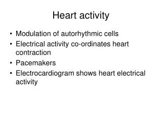
Heart activity • Changes in pressure and volume during the heart cycle
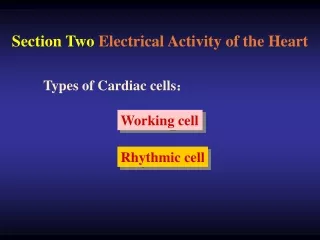
The heart cycle • Period from the beginning of one heart beat to the beginning of the next heart beat • Two phases: • Diastole, period of cardiac muscle relaxation • Systole, period of cardiac muscle contraction • The atria and ventricles do not contract and relax at the same time.
5 phases of the heart cycle • Heart at rest (atrial and ventricular diastole) • Completion of ventricular filling (atrial systole) • Early ventricular contraction (first heart sound) • The heart pumps (ventricular ejection) • Ventricular relaxation (second heart sound)
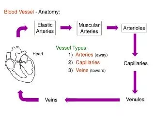
Heart at rest (atrial and ventricular diastole) • Atria and ventricles are relaxing • blood flows into the atria from veins • AV valves are open • Blood flows into the ventricles from the atria • Relaxed ventricles accept blood
2. Completion of ventricular filling (atrial systole) • Depolarization from the SA node • Contraction of atria • Blood pushed into the ventricles • Pressure increase accompanies contraction • Increase in pressure pushes some blood back to the veins
3. Early ventricular contraction (first heart sound) • Depolarization to AV node, down bundle of His, up Purkinje fibers • Ventricular systole begins at apex • Blood pushing up on AV pushes them shut • Blood does not flow back to atria • First heart sound when AV valves close (lub)
3. Early ventricular contraction (first heart sound) • The semilunar valves are also shut • Blood stays in the ventricle while it contracts • High pressure on the heart walls during this contraction • ISOMETRIC CONTRATION
3. Early ventricular contraction (first heart sound) • Atria begin to repolarize and relax Atrial pressure falls below venous pressure • Blood flows from veins to atria Blood stays in atria because the AV valves are closed
4. The heart pumps (ventricular ejection) • Ventricles contract • Semilunar valves open • Ventricular blood is pushed into the arteries
5. Ventricular relaxation (second heart sound) • Ventricular pressure falls • Blood starts to flow from arteries into ventricles • This backflow shuts the semilunar valves (dup of lub-dup) • Ventricles become closed • AV valves open when ventricular pressure is lower than atrial pressure
End Diastole Volume (EDV) • Ventricles are maximally filled at the end of ventricular relaxation (diastole) • When heart rate is very high, the ventricles may not have enough time to fill as much as when the heart rate is slow
End-systole volume (ESV) • The amount of blood left in the heart at the end of each contraction