Download
1 / 88
880 likes | 924 Vues
Explore the transmembrane potential, action potential phases, and ion channels of working and rhythmic cells in the heart.

E N D
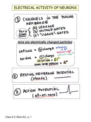
Section Two Electrical Activity of the Heart Types of Cardiac cells: Working cell Rhythmic cell
1. working cell: • including normal cardiac muscle cell ( atrial muscle and ventricular muscle) • having excitability, conductivity, contractility • no autorhythmicity
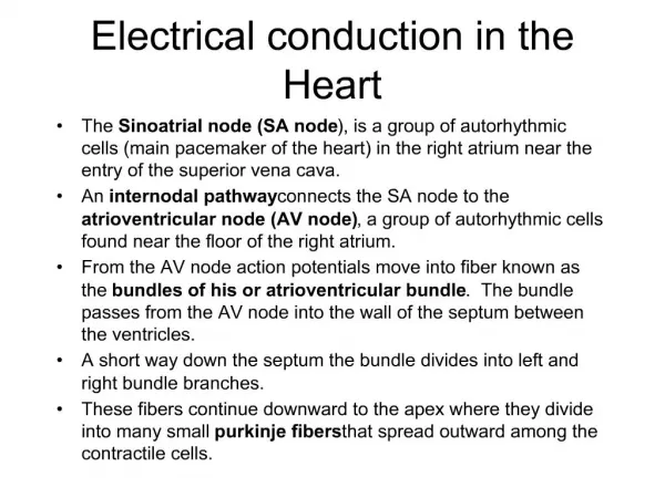
2. rhythmic cell:Specialized conduction system Sinoatrial node, Atrioventricular node, and Purkinje system. Special property: autorhythmicity
rhythmic cell: • mainly including pacemaker cell and purkinje cell . • having excitability, conductivity, autorhythmicity • no contractility
So cardiac muscle cell has four physiological properties. They are excitability, autorhythmicity, conductivity and contractility. But excitability, autorhythmicity and conductivity are called electrophysiological properties also.
Resting potential The resting membrane potential of individual cardiac muscle cells is about –90mV.
Ionic basis for RP and AP of working cell a. Ionic concentration differences across cell membrane b. Permeability to ions
Transmembrane potential of working cell ion intracellular extracellular equilibrium fluid fluid potential Na 30 140 +41 K 140 4 -94 Ca 0.0001 2 +132 Cl 30 104 -33 ( mmol/L ) ( mmol/L ) ( mV )
Na+ Cl- Cl- Cl- K+ K+ Na+ Cl- Cl- Na+ Na+ K+ K+ Na+ Cl- Cl- Cl- Cl- K+ Cl- K+ Cl- Na+ Cl- Cl- K+ Cl- Na+ Lipid bilayer membrane Na+ Cl- Cl- K+ A Cell 0 mV - X mV K+ Ik1 channel
Mechanism of RP genesis: Inward rectifier K+ channel Outward K+ current (Ik1) Na+ background current Na+ influx into cell Pump current:Na+ pump 3 Na+ out——2 K+ in
Basic concepts • Depolarization –– cations influx ---- Na+, Ca2+ inward current • Repolarization –– cations efflux ---- K+ outward current • Hyperpolarization: more negative than RP • Net current: inward > outward depolarization inward < outward repolarization inward = outward no change in Vm
Action potential Features : It lasts about 250-300ms, and can be divided into 5 phases. Depolarization proceeds rapidly, repolarization proceeds slowly.
+30 0 -50 Membrane potential (mv) -90 Time (ms)
Action potential (AP):300ms • (1) Phase o (rapid depolarization)1-2ms-90mv +30mv • (2)Phase 1(rapid initial repolarization) 10ms+30mv 0mv • (3)Phase 2 (plateau) 100-150ms 0mv • (4)Phase 3 (rapid late repolarization) 100-150ms 0mv -90mv • (5) Phase 4 (resting membrane potential) -90mv
Phase o : Stimulation ↓ partial depolarization ↓ threshold potential (-70mV) ↓ Na+ Ch. opening ↓ Na+ influx into cell Na+
Ionic basis for APPhase 0 (depolarization) Stimulation → partial depolarization (Na+ ) → threshold potential (-70mV) →Na+ Ch. opening →Na+ influx into cell down electrochemical gradient → less negative→0 mV → +30 mV (overshoot)
Features of fast Na+ channel (1). Activated and inactivated very fast. • Speed of depolarization: 200-400 V/s; • Fast channel • Fast response potential • Fast response cell
(2). Voltage dependent • Activation -70mV • Inactivation 0mV~+30mV • Recovery to reopen from -60mV • (3). Blocked by tetrodotoxin (TTX)
Regenerative process: • depolarization caused by Na+ influx • induces more Na+ channels to open and Na+ influx. • At the same time, K+ conductance falls and keeps depolarization state.
Phase 1 : Na+ Ch. is inactivated at +30mV + K+ Ch. is activated ↓ K+ outward current ↓ rapid repolarization ( Phase 1) 按任意键显示动画2 K+ Na+
Phase 1 (rapid repolarization) • (1) Na+ Ch. is inactivated at +30mV • (2) Transient outward current (Ito) K+ outward current, blocked by tetraethylammonium(TEA) and 4-aminopyridine.
Phase 2 : Ca2+ Ch. activation at –40mV + K+ Ch. is activated ↓ Inward Ca2+= outward K+ ↓ slow repolarization (Phase 2=plateau) 按任意键显示动画2 K+ K+ Na+ Ca2+
Phase 2 (plateau) Ca2+ Ch. activation at –40mV → Ca 2+ influx → Ca2+ inward current(L-type) IK Ch. is activated slowly at phase o –40mV • K+ slowly efflux → K+ outward current (延迟整流钾流)deactivated at phase 3 –50mV • Inward Ca2+ current = outward K+ current at early stage of plateau • Inward current < outward K+ current at late plateau, → more negative → repolarization
Close of IK1 Ch. at phase o and plateau prevents membrane potential from rapid repolarization • Phase 2 is the integration of inward Ca2+current and outward K+ current.
The features of Ca2+ channel: • (1).Slow channel, slow inward current, slow • activation and inactivation and • reactivation • (2).Voltage dependent: Activated at –40mV, inactivated at 0mV • (3).Blocked by Mn2+ and verapamil • (4).Low specialty: permeability to Na+ also.
Features of slow Ca2+channel • Slow channel • Slow response potential • Slow response cell • Two types L type -40mv blocked by Verapamil or Mn2+ T type -50mv blocked by Ni+.
Phase 3: Ca2+ channel is inactivated + K+ efflux via K+channel↑ ↓ rapid late repolarization ↓ RMP 3期 3期 ○ PUMP 按任意键显示动画2 Phase 4: Na+-K+ pump Na+-Ca2+exchange Ca2+ pump K+ K+ K+ ○ PUMP Na+ Ca2+
Phase 3 (late repolarization) • Ca2+ channel is inactivated. • ↑K+ efflux via IK channel • ↑K+ efflux via IK1 channel →↑outward • more and more K+ outward current↑→ • more and more negative → RMP.
Phase 4 (resting stage) • During Phase 0-3, Na+, Ca2+ and K+ imbalance outside and inside cell. • During Phase 4, Na+, Ca2+ efflux against concentration gradient; K+ influx against concentration gradient .
Na+-K+ pump: • 3 Na+ out and 2 K+ in • Na+-Ca2+ exchanger: • 1 Ca2+ out and 3 Na+ in • dependent of Na+ concentration difference inside and outside cell. • Ca2+ pump: Ca2+ out of cell. • Phase 4 (resting stage)
1 mV 2 0 3 0 4 - 90 Na+ + Ca2+ Na+-K+ K+ Ca2+ K+ K+ K+
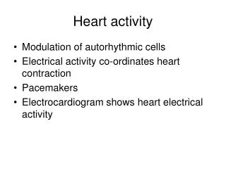
(Ⅱ) Transmembrane potential of Rhythmic cell Feature : phase 4 spontaneous depolarization the cause of autorhythmicity
Types of rhythmic cell • Autorhythmicity fast response cell –– Purkinje cell • Autorhythmicity slow response cell — S-A node and A-V node
1 P cells ① The level of maximal repolarization potential at the end of phase 3. (-70mv); threshold potential (-40mv). ② Phase 0 : I Ca-L, proceeds slowly,10V/s, 7ms, magnitude 70~85mV ③ No distinctphase 1 and phase 2. ④ Faster phase 4 spontaneous depolarization
phase 0 零电位 按任意键显示动画1、2 阈电位 Ca2+ (Ica-T) Ca2+ (Ica-L) phase 0 : phase 4 spontaneous depolarization→threshold potential (-40mv) → Ca2+ Ch. activation(Ica-L)→ Ca2+ influx
Features of slow Ca2+channel • Activated and inactivated very slow. • Slow channel • Slow response potential • Slow response cell • Two types L type -40mv blocked by Verapamil or Mn2+ T type -50mv blocked by Ni+
phase 3 按任意键显示动画1、2 K+ Phase 3:slow Ca2+ Ch(Ica-L)isinactivated + K+ Ch. is activated→ Ca2+ influx↓+ K+ efflux↑(IK)
phase 4 按任意键显示动画1、2 K+ If (Na+) Ca2+(ICaT) Phase 4:①IK declines and K+ efflux decreases* ②Inward current, If ↑(Na+) ③ ICaT largens and Ca2+ increases(ICa-T) → phase 4 spontaneous depolarization
a. Inward current, IfFeatures of If: (a) Carried by Na+, blocked by Cs, but not TTX (b) Activation at -60mV, full activation at –100mV (c) Noradrenalin → ↑If Acetylcholine →↓If
b. Inward Ca2+ current, ICa-T Activation at -50mV Blocked by Ni+ c. Gradually diminishing outward K+ current, Ik
LOOKING AT THE PACEMAKER CURRENTS voltage IK ICa-T If ionic currents ICa-L
2 Purkinje cell AP’s wave of Purkinje cell is similar to that of the working cells except phase 4.
Mechanism At phase 4, the membrane potential does not maintain at a level, but depolarizes automatically – the autorhythmicity • (Phase 0 – 3) Same as for ventricular cells • (Phase 4) PlusIf channels, depolarizesautomatically
Ionic basis of spontaneous phase 4 depolarization in cell-Purkinje cell (1) Gradual increase in inward current,If* (2) Gradually diminishing outward K+ current, IK If > IK , depolarization →threshold potential → a new AP
cardiac cells • Fast response, non –rhythmic cells: working cells • Fast response, rhythmic cells: cells in special conduction system of A-V bundle and Purkinje network.
3)Slow response, non-rhythmic cells: cells in nodal area 4)Slow response rhythmic cells: S-A node, atrionodal area (AN), nodal –His (NH) cells
Pacemaker cell Of SA node Ventricular working cell Purkinje cell