Cardiac electrical activity
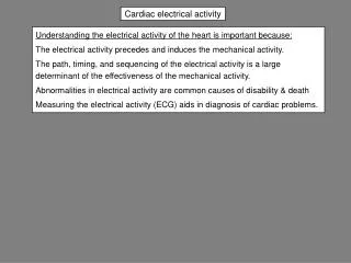
Cardiac electrical activity. Understanding the electrical activity of the heart is important because: The electrical activity precedes and induces the mechanical activity.

Cardiac electrical activity
E N D
Presentation Transcript
Cardiac electrical activity Understanding the electrical activity of the heart is important because: The electrical activity precedes and induces the mechanical activity. The path, timing, and sequencing of the electrical activity is a large determinant of the effectiveness of the mechanical activity. Abnormalities in electrical activity are common causes of disability & death Measuring the electrical activity (ECG) aids in diagnosis of cardiac problems.
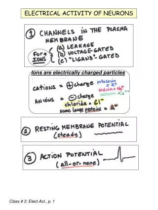
Electrical activity in biological systems is produced by diffusion of ions across membranes Ions diffuse across cell membranes through specific water-filled channels driven by the electric & concentration gradients across the membrane. In cardiac myocytes the resting membrane potential is produced mostly by diffusion of K+ down it’s concentration gradient out of the cell. Action potentials in nerve & muscle are due to rapid diffusion of Na+ into the cell down it’s electrochemical gradient. (Exception: action potentials in the SA and AV nodes are due to diffusion of Ca++ through L-type Ca++ channels.) Channels of interest in the cardiovascular system are: Voltage-gated: open with a change in membrane potential. Ligand gated: open in response to a hormone or intracellular signal. Stretch activated: open in response to stretch of a myocyte
Electrical gradient: Negative resting membrane potential drives K+ into cell. A - = intracellular anionic proteins & organic phosphates balance positive charge on K+. + + + + + + - - - - - - A- K+ = 4 mEq/L K+ = 135 mEq/L K+ = 4 mEq/L Concentration gradient: Higher cell [K+] drives K+ out of cell. Equilibrium potential (or Nernst potential): If the membrane potential = the equilibrium potential for an ion, the rates of passive diffusion of the ion into and out of the cell are equal. The equilibrium potential for an ion is the membrane potential at which the effects of electrical and concentration gradients on diffusion of the ion balance each other.
Na+ 10 inside outside 145 O/I = 14.5 K+ 135 inside I/O = 33.75 outside 4 Ca++ 0.0001 inside O/I = 20,000 2 outside Equilibrium potential (Nernst potential) for Na+, K+ & Ca++ Cardiac myocyte resting Membrane Potential = - 90 mV
Equilibrium potential compared to resting membrane potential (RMP)
Membrane pumps and potentials in a ventricular myocyte active [Na+] = 145 mEq/L RMP = -90 mV passive Na+ Na+ [Na+] = 10 mEq/L 3 Na+ 3 Na+ ATPase [K+] = 4 mEq/L 2 K+ 2 K+ [K+] = 135 mEq/L K+ K+ Sarcoplasmic reticulum: Ca++ store Ca++ released during excitation Ca++ taken up during relaxation by Ca++ATPase in SR (SERCA) Na+ Na+ Na + Ca++ exchange Ca++ Ca++ Ca++ Ca++ Ca++ATPase Ca++ Ca++ ATPase Ca++ Ca++ Voltage-gated L type Ca++ channel
0 Resting membrane potential -50 Transmembrane potential, mV -100 Equilibrium potential for K+ -150 1 2 3 5 10 20 30 50 [K+in] = 135 mEq/L Extracellular [K+], mEq/L The resting membrane potential is primarily determined by diffusion of K+ Changing extracellular [K+] changes the concentration gradient for diffusion of K+ out of the cell and the equilibrium potential (EK+, Nernst potential) for K+. Parallel changes in EK and the resting membrane potential mean that diffusion of K+ is the main determinant of the resting membrane potential.
+ + + + - - - - K+ = 135 mEq/L K+ = 4 mEq/L RMP = - 90 mV + + + + - - - - K+ = 135 mEq/L K+ = 7 mEq/L RMP = - 75 mV + + + + - - - - K+ = 135 mEq/L RMP = - 95 mV K+ = 2 mEq/L Changing extracellular [K+] changes the concentration gradient for diffusion of K+ out of the cell Normal Hyperkalemia: gradient for diffusion of K+ is decreased. Hypokalemia: gradient for diffusion of K+ is increased.
Chronic Acute Depolarization inactivates fast Na+ channels hyperkalemia depolarizes RMP excitability RMP approaches threshold excitability cardiac conduction velocity Skeletal muscle weakness Effects of acute and chronic hyperkalemia “Symptoms of hyperkalemia … do not become manifest until the plasma potassium concentration exceeds 7.0 mEq/L , unless the rise in potassium concentration has been very rapid.” (B.D. Rose, UpToDate 2008).
Acute Chronic Hypokalemia hyperpolarizes RMP Hyperpolarization activates some fast Na+ channels RMP becomes more negative relative to threshold excitability excitability cardiac arrhythmias Skeletal Muscle weakness Effects of acute and chronic hypokalemia Symptoms usually occur only if plasma K+ is below 2.5 to 3 mEq/L
2 1 zero mV 0 3 ARP RRP 4 - 90 mV 0 100 200 300 milliseconds Phases of a ventricular myocyte action potential (0, 1, 2, 3, 4) ARP = absolute refractory period RRP = relative refractory period
Ion channels in the cardiac myocyte action potential Plateau: L Type Ca + + channels open, Some K+ channels close • 40 mV: L-type • Ca++ channel activation 2 1 zero mV • 55 mV: fast Na+ channel inactivation Repolarization: Ca + + channels close, K+ channels open 3 0 • 70 mV upshoot: • threshold for fast Na+ channel activation 4 gNa+ - 90 mV gCa++ gK+ g = conductance = 1/resistance
1 2 3 0 4 Summary of ionic currents in cardiac myocyte action potential Influx: flow into cell. Efflux: flow out of cell
40 20 Upshoot of action potential 0 -20 Ventricular myocyte action potential Potential, mV -40 -60 Resting membrane potential -80 -100 15 30 50 150 Extracellular [Na+] The upshoot of the AP is determined by diffusion of Na+ into the cell As ECF [Na+] decreases (x axis) the magnitude (height) of the upshoot decreases (y axis) because the Na+ concentration gradient across the cell membrane decreases.
All or none law and force of contraction SR = sarcoplasmic reticulum
0 -20 -40 Threshold – 55 mV -60 -80 IK+ outward Ionic currents inward If ICa++ Pacemaker cells in the sino-atrial node set the normal heart rate The pacemaker potential is a spontaneously depolarizing membrane potential carried by a ”funny” Na+ current, If. This current is called “funny” because the responsible channels open as the cell membrane repolarizes (becomes more negative) past –50 mV. Other Na+ channels open when the membrane depolarizes. The upshoot of the action potential is a calcium current, ICa++. due to opening of L-type Ca++ channels. Repolarization is due to an outward K+ current, IK+ due to opening of K+ channels.
Path of excitation in the heart Sino-atrial node originates action potentials Atrial myocytes The heart is a syncytium. Depolarization (inward Na+ current) spreads from conducting tissue to myocytes & between myocytes via gap junctions. Gap junctions are located at the ends of myocytes and conduct current longitudinally. The velocity of conduction of APs is greatest in the Purkinje fibers. Atrioventricular node Bundle of His Right & left bundle branches Purkinje fibers The AV node is the only pathway for conduction between atria & ventricles Conduction is delayed in the AV node allowing optimal ventricular filling during atrial contraction. Ventricular myocytes
Conduction velocity Velocity of conduction of action potentials in cardiac myocytes and Purkinje fibers is directly proportional to: 1) Fiber diameter (greatest in Purkinje fibers). 2)The magnitude of the upshoot of the AP. 3) Rate of rise of the AP in phase 0 (increase slope of phase 0). Magnitude of AP Rate of rise of AP Local current (depolarization) Conduction velocity Both an increase in the magnitude of the action potential and a more rapid rate of rise of the AP in phase 0 cause greater depolarization of the cell membrane locally, opening more fast Na+ channels and increasing conduction,.
ischemia Activity of Na+. K+ ATPase Leak of K+ from cells extracellular [K+] K+ concentration gradient. Gradual depolarization potential driving Na+ entry Inactivation of some Na+ channels Magnitude of AP Rate of rise of AP Local currents Conduction velocity Conduction velocity, plasma [K+] & ischemia Depolarization from any cause decreases conduction velocity in Purkinje fibers & myocytes. Conduction velocity in hyperkalemia leads to abnormal conduction & disturbances in cardiac rhythm.
Automaticity & latent pacemakers In addition to the SA node, the AV node & Purkinje fibers may show pacemaker potentials. The heart rate is set by the pacemaker that is depolarizing at the fastest rate, normally the SA node. Damage or blockade of the SA node will allow slower pacemakers to take over & results in bradycardia (decreased HR).