Download
1 / 40
400 likes | 634 Vues
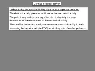
Electrical Treatment for Cardiac Abnormalities. Advanced Paramedic Skills Mary Osinga. Objectives. Defibrillation Theory What gets defibrillated Safety review Placement AED Introduction Pacing Overview Transcutaneous Implanted AED’s. The AHA Chain of Survival.

E N D
Electrical Treatment for Cardiac Abnormalities Advanced Paramedic Skills Mary Osinga
Objectives • Defibrillation • Theory • What gets defibrillated • Safety review • Placement • AED Introduction • Pacing Overview • Transcutaneous • Implanted AED’s
The AHA Chain of Survival 1. Early access to the emergency medical services (EMS) system 2. Early CPR either by bystanders or first-responder rescuers 3. Early defibrillation by first responders, emergency medical technicians (EMTs), paramedics, or nurses and physicians if they are on the scene 4. Early ACLS Source: Cummins et al., 1991
Chain of Survival- Purpose • EARLY ACCESS • to911 system. To get medics moving. • EARLY CPR • to help circulated oxygen to the patient's heart and brain. • EARLY DEFIBRILLATION • May be AED on scene, such as health clubs, fd etc • shocks to restore normal heart rhythm. • EARLY ADVANCED CARE • provided by als or hospital staff.
Most survivors of cardiac arrest are from the group of patients . . . • Whose collapse is witnessed by a bystander, • Who receive cardiopulmonary resuscitation (CPR) within 4 to 5 minutes, and • Who receive advanced cardiac life support (ACLS), e.g., defibrillation, intubation, drug therapy, within the first 10 minutes. Source: Weaver et al., 1986
Survival Rates No CPR 0%-2% surviveDelayed defibrillation Early CPR 2%-8% surviveDelayed defibrillation Early CPR 20% survive Early defibrillation Early CPR 30% survive Very early defibrillation Early ACLS Source: American Heart Association, 1994
Minutes elapsed 1 2 3 4 5 6 7 8 9 10 11 Defibrillation Statistics • Defibrillations chances of restoring a pulse decrease rapidly with time.
AHA says… • Most frequent initial rhythm in SCD is VF • ONLY effective treatment is defibrillation • Probability of successful conversion diminishes over time • Speed at which defib shock is delivery is MAJOR determining factor
Need for Defibrillation? • Only put the unit on someone you would do CPR on... someone who is • Unresponsive • Not breathing • and has NO signs of circulation or no pulse. • I.e do the LOC, ABC’s first 11
Always Start with Basics • First paramedic- • Assess responsiveness • Airway, no air in and out – oral airway in • Breathing – none –start bagging • Circulation-none- landmark and start CPR • Second Medic • Gets out defibrillator, sets up • Attaches big pads • Works monitor
Ventricular Fibrillation • Ventricular fibrillation (VF) is an abnormal heart rhythm often seen in sudden cardiac arrest. • This rhythm is caused by an abnormal and very fast electrical activity in the heart. • VF is chaotic and unorganized; the heart just quivers and cannot effectively pump blood. • There IS electrical activity but No mechanical pumping
Ventricular Fibrillation • VF will be short lived and will deteriorate to asystole if not treated promptly. • For each minute that VF persists, the likelihood of successful resuscitation decreases by approximately 10 percent.
Ventricular Fibrillation • Ventricular fibrillation (VF) is an abnormal heart rhythm often seen in sudden cardiac arrest. • This rhythm is caused by an abnormal and very fast electrical activity in the heart. • VF is chaotic and unorganized; the heart just quivers and cannot effectively pump blood.
Ventricular Fibrillation This rhythm can be coarse or fine, (close to asystole) 16
Ventricular Tachycardia VT This rhythm is wide complex (greater than…?) No discernable P or T waves 17
Defibrillation Theory • Definition-the process of passing a current through the fibrillating heart to depolarize the cells and allow for repolarization by a pacemaker cell • Need to shock a critical mass of myocardium • Otherwise ectopi foci remain fibrillating
Defib theory continued • Defibrillator is a capacitor that stores NRG • Consists of capacitor, high voltage power supply and delivery conduits (pads or paddles) • Various waveforms of NRG, such as monophasic and biphasic (less NRG required) • Use predominately DC • NRG=Power x duration • Joules =watts (not WHAT’s) x Seconds • Resistance to defibrillation success are:
Resistance in Chest Wall to J’s • Paddle or pad pressure • Pad-skin contact (hair etc) • Pad-paddle skin surface area • Number of previous countershocks • Concept of transthorasic impedance • Time of respiratory cycle (ideally inspiratory)
Success of defibrillation • Time from onset of chaotic rhythm • Condition of myocardium • Heart size and body weight • Impedance • Pad size • Placement • Interface • Defibrillator working and delivering proper energy setting
General Considerations • Wet patients (drowning etc) • Medication patches • Implanted pacemakers • Young patients • Excessive chest hair 22
Patient's Clothing • The chest should be exposed to allow placement of the disposabledefibrillation electrodes. • Clothes may need to be cut with shears to facilitate early defibrillation.
Defibrillation = Unsynchronized Cardioversion • Used exclusively as the definitive treatment for ventricular fibrillation and pulseless ventricular tachycardia • A energy used to settle a chaotic heart rhythm temporary into asystole, in the hopes that some pacemaker cell in the heart will start an organized rhythm. • Start with 200J, then 300J and 360J
Steps for Defibrillation • Ensure pulselessness (longer pulse checks for hypothermic patients) • Hook up either hands-free pads or paddles to chest with gel pads. • Start CPR (May do basic airway and vent, but do not delay defibrillation for these maneuvers) • Press Analyze • If vfib or pulseless V tach- machine will say “stand clear” • monitor charges to preset voltage( to 200 J) • ensure no one touching patient including you • Defibrillate at 200Joules with LP 12 or other defibrillator • Do not touch patient • Reanalyze and repeat at higher J settings 300 • Reanalyze – still vfib/vtach charge to 360J and press shock • Once at 360, stay at that setting.
Defib Pad Placement • Attach anterior pad to R shoulder below the clavicle R of the sternum • Lateral pad is anterior axillary line at the level of the base or apex of heart -ensure good contact- shave if required
Defibrillation • Must be 25 lbs pressure with paddles to ensure good contact and success of defibrillation • Stacked shocks in beginning 200/300/360J are to decrease transthorasic resistance. If you take too long between shock, this is less effective • Can also defib anterior/posterior but more difficult and cumbersome in the VSA patient
AED Standing Order Review • Shockable rhythms
AED Standing Order Review for Non-shockable rhythms • Asystole • Anything else with no pulse = PEA or pulseless elctrical activity
Cardioversion= Synchronized • Used for unstable patients in supraventricular and ventricular fast rhythms with a pulse, in order to slow them down • Rhythms like SVT, rapid Afib/flutter, Vtach, PSVT • Pad placement is the same as for defibrillation • ENSURE THAT WHEN YOU DO THIS, YOU PRESS THE ‘SYNC’ BUTTON ON MONITOR!!
Symptomatic Tachyarrhythmias • Look for these signs/symptoms before aggressively electrically treating a patient • There is no rule on which or how many signs a patient needs to have to be treated electrically, use experience and judgement if no patch available • Chest pain • Shortness of breath • Pulmonary edema • Altered LOC • Hypotension • Syncope • diaphoresis
Find it on your monitor! What does the ‘SYNC” button do? • This identifies the R waves on the ECG and marks them (will see a ‘tag’ on them) • This tells the machine what timing to use in order to identify the absolute refractory period • Do NOT want to cardiovert at this time! • What will happen if you do? (if the machine failed to sense this or worse, YOU failed to press the ‘sync’ button before you shocked?
!!! This is bad • Yes indeedy….you could put them into vfib • you took a organized rhythm and shocked during the absolute refractory period (R on T ) kind of thing and produced a BADDDD thing! • Always double check before shocking that sync is ‘on’ • NOTE: most defibs (LP12 included) have an automatic ‘sync’ shutoff in case patients go into vfib anyway. SO make sure you press it in before EACH cardioversion!
Some info for Paramedic • Again, defibrillation may be interfered with by other equipment • Notify partner/other helpers of procedure • Watch for skin burns • Remove NTG patch • Ideally, do not have O2 nearby! • Ensure everyone clear when you defib!
Contraindications • No order for it! • Severe hypothermia-reduced algorithm • Code 5 Patient • Open chest wounds • In a wet environment Rule of thumb: If patients says “what are you doing?” you do not need to defibrillate!
Transcutaneous Pacing • For symptomatic bradycardias • examples are anything from sinus bradycardia (rare) to 2nd degree type I and II and Third degree block • If it needs speeding up, you could potentially pace it. • May also attempt to pace asystole or slow idioventricular VSA if arrest is new and pacer is quickly available • Standby pacing (pads on but not actually pacing) is indicated for patients in 2nd degree Type II or third degree who are stable
Procedure for Pacing • Explain to patient what you are doing • IV , O2, ECG (and backup airway equipment) • Sedate as indicated from BHP • Attach pads to patient. Ideally anterior/posterior (sandwich) is best for contact and success. Anterior pad over left lower hemithorax. Posterior in the subclavicular area with superior margin just below the clavicles. Good contact is essential • Connect cables to LP 12 • Set demand (turn pacer to ‘on” • set HR (between 60-80) • start increasing mA from O until get capture on screen • ensure pulse matches monitor • add 10 mA to ensure safe zone • Check vitals (pulse, BP and mentation) • recheck for capture periodically
Community AED’s • More and more people trained to use AED’s, fully automated versions • Know models of defib and know how to get report (what happened?) • Give rescuers good feedback during transfer of care
Where to place AEDs? • In a medical clinic (if available). • In a reception or common area. • Near a fire extinguisher. • With a safety response team member. • With a security officer. • On board anairline jet. • AEDs should be visible and easily accessible.
For Next Week • Please read defibrillation and cardiac monitoring in book • ECG monitoring pgs 1206-1271 (hopefully review) • Defibrillation pgs 1297-1305 • PLEASE READ ABOVE FOR SURE!