Heart
Discover the intricate details of the heart's anatomy, functions, and circulatory system. Learn about cardiac membranes, chambers, valves, and coronary circulation. Get insights into the cardiac cycle and cardiac output dynamics.

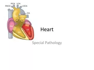
Heart
E N D
Presentation Transcript
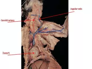
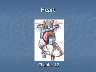
Heart The heart pumps blood, which creates pressure, and circulates oxygen, nutrients, and other substances.
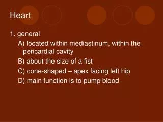
Heart Information • Cardiology is the study of the heart • In the embryo, the heart begins to beat at 4 weeks • The heart beats 100,000 times a day • The heart is located in the thoracic cavity between the lungs, and above the diaphragm.
Cont. • Is located mediastinally within chest • The location makes CPR possible • Is about the size of the person’s closed fist • The heart is composed of cardiac muscle
Pericardial Membranes The pericardium is a triple layered sac that surrounds and protects the heart 2 portions • Fibrous- (pericardial sac) tough, loose fitting sac that prevents stretching, provides protection • Serous- delicate membrane that forms double layers around heart a. parietal b. epicardium
Pericardial Membranes • Serous fluid, which prevents friction as the heart beats, is between the parietal and epicardium layers of pericardium • Pericarditis is the inflammation of this fluid
Layers of the heart • Fibrous • Parietal • Epicardium- outermost • Myocardium- responsible for pumping (heart muscle) • Endocardium- provides smooth lining for inside of heart
4 chambers of the heart • Right atrium • Left atrium • Right ventricle • Left ventricle The two superior chambers are the left/right atrium The two inferior chambers are the left/right ventricle
Cont. • Auricle- are found on each atria and increase size and volume (ear shaped) • Coronary sulcus- are external grooves that separate the atria from the ventricles, and contain coronary blood vessels
Myocardium is the muscle of the heart • The chambers are lined with endocardium, which is a smooth tissue that prevents abnormal blood clotting
The interatrial septum is an internal wall that separates the atria • The interventricular septum is an internal wall that separates the ventricles • Fossa ovalis- remnant of fetal heart opening found in right atrium
Right Atrium • Receives blood from the upper body by way of the superior vena cava and receives blood from the lower body by way of the inferior vena cava. • The tricuspid valve prevents the backflow of blood into the right atrium
Right ventricle • When the right ventricle contracts, the tricuspid valve closes, and the blood is pumped to the lungs through the pulmonary artery • The flaps of the pulmonary semilunar valve close to prevent the backflow of blood into the right ventricle
Right ventricle • The right ventricle has columns of myocardium(muscle) called papillary muscles. • The chordea tendineae, extend from the papillary muscle to the flaps of the tricuspid • The chordea tendineae prevent the inversion of the tricuspid valve
Left atrium • The left atrium receives blood from the lungs, by way of four pulmonary veins. • This blood then flows into the left ventricle through the bicuspid valve • The bicuspid valve prevents backflow of blood from the left ventricle to the left atrium when the left ventricle contracts. • Another function is to produce the hormone ANH (atrial natriuretic hormone) which lowers blood pressure
Left ventricle • The walls of the left ventricle are thicker than those of the right ventricle, which enables the left ventricle to contract more forcefully. • The left ventricle pumps blood to the body through the aorta, the largest artery of the body.
Left ventricle • The chordae tendineae anchor the bicuspid valve of the left ventricle • At the junction of the aorta and the left ventricle is the aortic semilunar vavle. • The aortic semilunar valve closes when the left ventricle relaxes, to prevent the backflow of blood from the aorta to the left ventricle
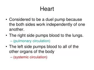
Heart summary • The heart is really a double, or two sided pump. • The right side of the heart receives deoxygenated blood from the body and pumps it to the lungs to pick up oxygen and release carbon dioxide. • The left side of the heart receives oxygenated blood from the lungs and pumps it to the body. • Both pumps work simultaneously, both atria contract together, followed by the contraction of both ventricles.
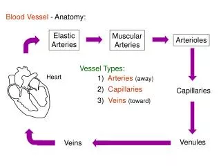
Coronary circulation • Coronary circulation is the flow of blood through the vessels of the myocardium • Coronary arteries supply blood • Coronary veins drain (greater, middle) • Ischemia- when part of the myocardium is deprived of blood
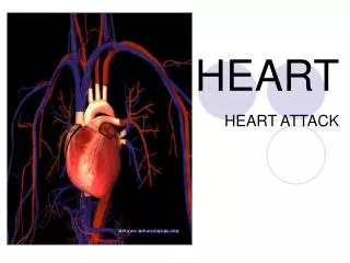
Coronary vessels • Prolonged ischemia will create an infarct, and an area of necrotic (dead) tissue. Commonly known as a heart attack.
Cardiac OutPut • Cardiac Output is the amount of blood pumped by a ventricle in 1 minute • Stroke Volume is the amount of blood pumped per beat • Starling’s Law of the Heart states that the more cardiac muscles are stretched, the more forcefully they contract
Cardiac cycle • Cardiac cycle is the simultaneous contraction of the two atria, followed .20 second later by the simultaneous contraction of the ventricles. • 1% of cardiac muscle fibers become autorhythmic cells (generate action potentials) • These cells form the conduction system for the heart
Cardiac Conduction pathway • SA node- found in right atria • AV node- found in atria septum • Bundle of His- found between atria and ventricles • Purkinje fibers- found in apex of ventricles
Cardiac cycle • Systole is another term for contraction. • Diastole is another term for relaxation.
Heart sounds • Each heartbeat produces four sounds, two are heard lubb-dupp • The first sound lub, the loudest and longest, is caused by the closing of the AV valves. • The second sound dup, is caused by the closure of the aortic and pulmonary semilunar valves.
Heart sounds • If any valves do not close properly, an extra sound called a heart murmur may be heard.
Heart rate • A healthy adult has a resting heart rate (pulse) of 60 to 80 beats per minute, which is the rate of depolarization of the SA node. • A rate of less than 60 (except for athletes) is called bradycardia; a rate greater than 100 beats is called tachycardia.
Heart rate • A child’s normal heart rate may be as high as 100, that of a infant may be as high as 120, and that of a near term fetus as high of a 140 beats per minute. • These higher rates are not related to age, but to rather to size; the smaller the individual the faster the rate.
Regulation of heart rate • The medulla of the brain regulates the heart rate; it contains the two cardiac centers, the accelerator, and the inhibitory center. • Pressoreceptors in the aortic sinus detect changes in blood pressure. • Chemoreceptors in the aortic body detect changes in the oxygen content of the blood.
Electrocardiogram • A ECG is a recording of action potentials produced by the myocardium during each heart beat • A ECG consist of three waves • P wave-small upward wave, that represents atrial depolarization • QRS complex- represents the depolarization of the ventricles - large Q indicate myocardial infarction - enlarged R indicates enlarged ventricles - flatter T indicates insufficient O2 to heart muscle
ECG 3. T wave- represents the repolarization of the ventricles.
Risk Factors in Heart Disease • Smoking- Nicotine enters blood stream and constricts small blood vessels, which elevates pressure • Obesity- have extra capillaries in adipose tissue (200 mi), more work • High blood pressure- causes the left ventricle to enlarge
Treatments • Hypothermia- slows metabolism and reduces needs of tissue • Heart transplant- 50 to 1 • Cardiac assist - Cardiomyoplasty- wrapping heart in muscle - LVAD- implanted in abdomen - Hemopump- propeller like pump
Aging and the heart • Heart muscle becomes less efficient with age. • Atherosclerosis, hypertension, and arrhythmias become more common.
Astherosclerosis • Areas where there are a build up cholesterol plaques in the coronary artery. These areas narrow the artery and increase the risk of clot formation, reducing the flow of oxygen. • Predisposing risk are cigarette smoking, diabetes mellitus, and high blood pressure (genetics). • Can be treated with angioplasty or bypass
Arrhythmias • Are irregular heartbeats caused by damage to part of the conduction pathway. • Palpitations are non-serious irregular heartbeats, and may result from too much caffeine, nicotine, or alcohol. • Flutter is a very rapid but fairly regular heartbeat. • Fibrillation is a very rapid and uncoordinated ventricular beat that is totally ineffective for pumping blood (V-fib)
Rheumatic Fever • An infection caused by Streptococcus bacteria that inflames the heart damaging the valves by stretching