Heart
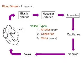
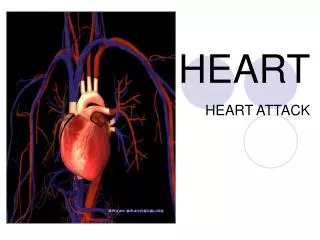
Heart. Chapter 12. Heart System of a pump and pathway for blood to travel Arteries - vessels that carry blood a way from the heart Veins - vessels that carry blood to the heart Capillaries are very small vessels that connect the two at the microscopic level. Exchange nutrients for waste

Heart
E N D
Presentation Transcript
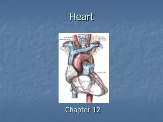
Heart Chapter 12
Heart System of a pump and pathway for blood to travel Arteries- vessels that carry blood away from the heart Veins- vessels that carry blood to the heart Capillaries are very small vessels that connect the two at the microscopic level
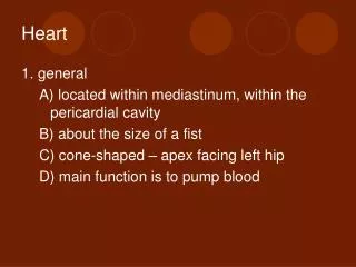
Exchange nutrients for waste • Exchange O2 for CO2 Heart lies in the pericardial cavityin the lower portion of the mediastinumresting on the diaphragm. The inferior portion has a point or tip called the apex. Broad upper surface called the base
The pericardium consists of two principle portions. 1- fibrous pericardium 2- serous pericardium Fibrous pericardium: tough and inelastic - rests on and attaches to diaphragm. - provides protection
Serous pericardium: - thinner, delicate, double layered. • 1- Visceral pericardium: (epicardium) contacts the surface of the heart • 2- Parietal pericardium: fused to the fibrous pericardium • Pericardial cavity: separates two layers with pericardial fluid (lubricates membranes) - cardiac tamponade
Heart consists of three layers: 1- Endocardium: lines inside of heart and heart valves 2- Myocardium: thick heart muscle. Blood supplied by coronary arteries. involuntary, striated, intercalated disks 3- Epicardium: smooth outer layer
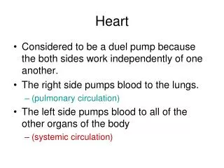
Heart is a double pump Right side of the heart is the receiving side. - pulmonary circulation Left side of the heart is the giving side. - systemic circulation (the body)
Heart has four chambers Two upper chambers are called atria (sing. atrium) Right and left Two lower chambers called ventricles Right and left Right and left ventricles are separated by a thick walled structure called the Interventricular septum
Blood flow between chambers and larger arteries pass through valves. Valves that connect the upper atria to the lower ventricles are termed atrioventricular valves (AV). There are two 1- Tricuspid valve. This “3 flapped” valve connects the right atria to the right ventricle 2- Bicuspid valve (mitral): This “2 flapped” valve connect the left atria to the left ventricle
Valves that connect the lower ventricles to the larger arteries carrying blood away from the heart are called semilunar valves. 1- Pulmonary semilunar valve: Valve located between the right ventricle and the pulmonary artery 2- Aortic semilunar valve: Valve located between left ventricle and aorta.
Contraction of the heart is systole Relaxation of the heart is called diastole When the heart beats the atria contract first and together then the ventricles contract second and together. Deoxygenated blood drains into right atrium from - superior vena cava - inferior vena cava - coronary sinus (from heart)
Blood is supplied to the heart by the first two branches of the aorta, right and left coronary arteries. These arteries arise from the base of the aorta and encircle the heart in the atrioventricular groove. • Left coronary artery: • anterior interventricular artery - supplies blood to the interventricular septum and anterior walls of both ventricles • circumflex artery - supplies blood to the left atrium and the posterior walls of the left ventricle
Right coronary artery: • Posterior interventricular artery - runs to the apex and supplies blood to the posterior ventricular walls • Marginal artery - supplies blood to the myocardium of the right side of the heart • The myocardium needs a constant supply of oxygen in order for the heart to continually pump. Myocardium capillaries are branches of cardiac veins which join to form the coronary sinus, an enlarged vein which empties into the right atrium.
Coronary veins: -Great cardiac vein, -Middle cardiac vein, -Small cardiac vein… … drains into cardiac sinus which drains into right atrium.
Conduction and Pacemaker Conductility- ability to conduct an electrical impulse These impulses must be coordinated. Intercalated disks are electrical connectors that join the muscle fibers. Remember: Atria contract together then ventricles.
1- Sinoatrial node (SA node) pacemaker of the heart. 2- Atrioventricular (AV node) 3- Bundle of His- interventricular septum - right and left bundle branches 4- Purkinje fibers- ventricle walls
Cardiovascular (CV) center is located in the medulla of the brainstem. • Sympathetic nervous system: cardiac accelerator nerves coming off of thoracic spinal cord segments. Increases heart rate • Parasympathetic nervous system: Vagus nerve (CNX) coming off of medulla (brainstem). Decreases heart rate.
ELECTROCARDIOGRAM (ECG or EKG) (Action Potential) Conduction system can be measured P QRS T