Heart
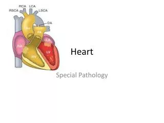
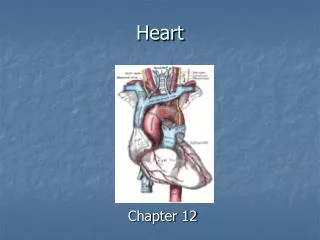
Heart. Anatomy of the Heart with 2D animation Note: by moving the mouse over the heart students will be able to hear the pronunciations. http://www.nucleuscatalog.com/cardiovascular-system/search?search_category=3556.

Heart
E N D
Presentation Transcript
Heart Anatomy of the Heart with 2D animationNote: by moving the mouse over the heart students will be able to hear the pronunciations.http://www.nucleuscatalog.com/cardiovascular-system/search?search_category=3556
In a particular hospital’s Intensive Care Unit, patients always died in the same bed, on Sunday morning, at about 11:00 a.m., regardless of their medical condition. This puzzled the doctors and some even thought it had something to do with the supernatural. No one could solve the mystery as to why the deaths occurred around 11:00 a.m. on Sunday, so a Worldwide team of experts was assembled to investigate the cause of the incidents. The next Sunday morning, a few minutes before 11:00 a.m., all of the doctors and nurses nervously waited outside the ward to see for themselves what the terrible phenomenon was all about. Some were holding wooden crosses, prayer books, and other holy objects to ward off the evil spirits. Just when the clock struck 11:00, Pookie Johnson, the part-time Sunday sweeper, entered the ward and unplugged the life Support system so he could use the vacuum cleaner.
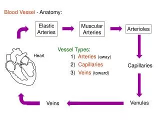
CIRCULATORY SYSTEMFUNCTIONS: • Transport (nutrients, wastes, oxygen, CO2, hormones) • Immunity (leukocytes, antibodies) • Temperature regulation (when you are cold, blood vessels constrict; when hot, they dilate) • Penile erection
COMPONENTS OF CIRCULATORY SYSTEM • Heart • Blood • Blood vessels (arteries, capillaries, veins) • Lymph vessels and nodes
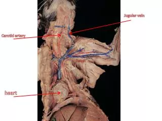
THE HEART • The heart is the simplest organ in the body. It does only one thing: pumps blood. • It beats 42 million times a year. • It’s about the size of your clenched fist. Some of you have big fists, some have smaller fists. • Its location is deep to the sternum. Take your left fist and place it on the sternum, then angle the bottom of your wrist to the left. When you say the Pledge of Allegiance, your hand is not over your heart. It’s not on the left, it’s in the center.
Location of the Heart in the Thorax The heart is located in the MEDIASTINUM, which is the central compartment of the thoracic cavity.
Layers of tissues around the heart: PERICARDIUM 1) Parietal pericardium 2) Pericardial cavity 3) Visceral pericardium HEART 1) Epicardium (same as visceral pericardium) 2) Myocardium 3) Endocardium Visceral pericardium Parietal pericardium
PERICARDIUM Surrounds the heart (like a heart in baggie). The function is to lubricate the heart, so as it beats, it won’t rub against anything. • The pericardium is divided into two layers with a space between them filled with fluid: • PARIETAL PERICARDIUM • PERICARDIAL CAVITY • VISCERAL PERICARDIUM
PARIETAL PERICARDIUM This is actually the outermost layer of thoracic cavity. Two layers: • SEROUS LAYER (simple squamous epithelium). Watery fluid. • FIBROUS LAYER (moderately dense fibrous connective tissue)
VISCERAL PERICARDIUM (aka EPICARDIUM) Outermost layer of heart. It also has two layers: • SEROUS LAYER • FIBROUS LAYER
Layers of tissues around the heart: PERICARDIUM 1) Parietal pericardium 2) Pericardial cavity 3) Visceral pericardium HEART 1) Epicardium (same as visceral pericardium) 2) Myocardium 3) Endocardium Visceral pericardium Parietal pericardium
Structure of the Heart Parietal pericardium Pericardial cavity Visceral pericardium Myocardium of heart Endocardium of heart Inside chamber of heart Figure 18.3
MYOCARDIUM The heart muscle itself (myocardium) is made of what tissue? Cardiac muscle.
ENDOCARDIUM • The lining on the inside of the heart. Has two layers: • ENDOTHELIUM (simple squamous epithelium that provides a smooth surface for the blood to pass by). • If there are any irregularities in the smoothness of the endothelium, a platelet could catch on it and start a blood clot. • Loose connective tissue (deep to the endothelium)
PERICARDITIS • Inflamed outer layer of heart. • Fluid accumulates in pericardial cavity, putting pressure on heart. • Pericarditis can also be caused by damage to the blood vessels blood leaks into pericardial cavity pressure. • Pericarditis can lead to pericardial friction rub, adhesions, and additional excess fluid in the pericardial cavity. It does NOT lead to a myocardial infarct (heart attack).
PERICARDITIS • CARDIAC TAPENADE: In severe cases of pericarditis, for example if there is a stab wound to the heart wall that causes fluid to exude into the pericardial cavity so much that the excess fluid compresses the heart and diminishes the heart’s ability to pump and it causes an irregular heart beat. An irregular heart beat is called arrhythmia. But Don’t get this confused with arrhythmia that is caused from a problem with the SA or AV node. The irregular heart beat from cardiac tapenade is caused from fluid entering the pericardial cavity and putting pressure on the heart. • Treatment is to stick a needle in the pericardial cavity and drain the fluid. http://catalog.nucleusinc.com/generateexhibit.php?ID=68030&ExhibitKeywordsRaw=&TL=&A=2
ENDOCARDITIS • More serious: • Bacteria enter bloodstream from a break in the skin (dental procedures, IV drug abuse, catheter) damage to lining and valves blood clots. • Those who already have damaged heart valves need prophylactic antibiotics. • Don’t get endocarditis (bacterial infection) mixed up with pericarditis, which can lead to cardiac tapenade.
THE HEART IS TWO PUMPS: LEFT AND RIGHT LEFT PUMP: From lungs to body RIGHT PUMP: From body to lungs
The Pulmonary and Systemic Circuits Figure 18.1
Heart Chambers • Each pump has two types of chambers: ATRIUM and VENTRICLE Left Atrium Right Atrium Right Ventricle Left Ventricle
Blood Flow • Deoxygenated blood from body enters the RA through the superior and inferior vena cava. • It pours through the TRICUSPID (RIGHT AV) VALVE into the right ventricle. • Right atrium contracts, pushes blood into the right ventricle ventricle expands, then contracts with force. • To prevent the blood from going back up into the atrium, need a valve.
Blood Flow • Blood comes from the superior or inferior vena cava, into the right atrium, past the tricuspid valve, into the right ventricle. It then goes past the pulmonary semilunar valve, and into the pulmonary arteries. Pulmonary arteries SVC RA Pulmonary Semilunar valve Tricuspid valve RV IVC
VALVES • Valves are like a swinging door that can only open one direction. But you can push against this door, since it’s only tissue. • But if you tie a rope to the doorknob, it won’t be able to go the wrong way. The ropes are called CHORDAE TENDONAE, (“heart strings”) which are anchored to pieces of myocardium called PAPILLARY MUSCLES. • The contraction of the papillary muscles pulls on the chordae tendonae to close the valves, preventing a PROLAPSED VALVE (turned inside out).
Valves Figure 18.9a
Function of the Atrioventricular Valves Figure 18.9b
Heart Valves – Valve Structure Figure 18.8a
Trabeculae carneae (spongy meat) • Trabeculae carnae are masses of irregular spongy tissues which project from the inner surface of the right and left ventricles. • The purpose of the trabeculae carneae is to prevent suction that would occur with a flat surface and thus impair the heart's ability to pump efficiently. • The trabeculae carneae also serve a similar function to papillary muscles in that their contraction pulls on the chordae tendineae, preventing prolapsed mitral (bicuspid) and tricuspid valves.
What is an artery? • An artery is a vessel that carries blood AWAY from the heart. It does not matter if it is oxygenated or deoxygenated blood. • A vein is a vessel that carries blood TOWARD the heart. It does not matter if it is oxygenated or deoxygenated blood.
Blood Flow With the ventricular contraction, blood can go only one way: into the PULMONARY ARTERY (one of the few arteries with deoxy blood). When the ventricles relax, the PULMONARY SEMILUNAR VALVE closes to prevent blood from going from the pulmonary artery back into the right ventricle. Pulmonary arteries SVC Pulmonary veins RA Pulmonary semilunar valve Tricuspid valve RV IVC
Function of the Semilunar Valves Figure 18.10a, b
Blood Flow • When the ventricles relax, the PULMONARY SEMILUNAR VALVE closes to prevent blood from going from the pulmonary artery back into the right ventricle. • Do the semilunar valves have a chordae tendonae? • No; the blood is not being forced back (with a contraction), it just falls back with gravity, so there’s not as much pressure.
Blood Flow • Blood then goes into lungs, gets oxygenated, and returns on the left side through the PULMONARY VEINS (one of the few veins with oxy blood), into the LEFT ATRIUM. Lungs Pulmonary arteries Pulmonary veins LA Bicuspid (mitral) valve Tricuspid valve LV
Blood Flow From the LEFT ATRIUM, it goes through the MITRAL VALVE (BICUSPID VALVE) into the LEFT VENTRICLE(there are also chordae tendonae here), which contracts. Therefore, the left ventricle is the chamber which is responsible for generating the largest pressure upon contraction. Lungs Pulmonary arteries Pulmonary veins LA Bicuspid (mitral) valve Tricuspid valve LV
Mitral Valve • The mitral valve gets its name from being the shape of a Bishop’s hat, called a mitre.
Blood Flow • The blood then goes past the AORTIC SEMILUNAR VALVE, into the AORTA, and out to the body. Aorta The semilunar valves are located between the ventricles and the great arteries. Pulmonary arteries SVC Pulmonary veins LA RA Pulmonary semilunar valve Bicuspid valve Tricuspid valve RV LV IVC
VALVES: “Try before you Buy” • TRICUSPID VALVE • BICUSPID VALVE (MITRAL VALVE) • PULMONARY SEMILUNAR VALVE • AORTIC SEMILUNAR VALVE • Structures associated with tricuspid and bicuspid valves • CHORDAE TENDONAE • PAPILLARY MUSCLES
Blood Flow Deoxy blood sup/inf vena cava R atrium tricuspid valve R ventricle pulmonary semilunar valve pulmonary artery lungs pulmonary veins Left atrium mitral (bicuspid) valve Left ventricle aortic semilunar valve aorta rest of body. Aorta Pulmonary arteries SVC Pulmonary veins LA RA Pulmonary semilunar valve Bicuspid valve Tricuspid valve Aortic semilunar valve RV LV IVC
Heart Chambers Figure 18.5e
Heart Chambers Figure 18.5b
Inferior View of the Heart Figure 18.5d
Atrial septum Right atrium Left atrium Left ventricle Right ventricle Apex Interventricular septum
Aortic semilunar valve Mitral (bicuspid) valve Cordaetendonae Papillary muscles Trabeculaecarnae
Mitral Valve Stenosis • If there is stenosis (blockage) of the mitral valve, where will the blood back up into? • Answer: the pulmonary circulation. Lungs Pulmonary arteries Pulmonary veins LA Bicuspid (mitral) valve Tricuspid valve LV
HEART BEATS • The pressure of blood against blood vessel walls is called blood pressure. • Blood pressure is recorded systole over diastole. Normal resting blood pressure is said to be 120/80. When blood pressure is too high, it is called HYPERTENSION. • The sound your heart makes when it is beating is the sound of the blood hitting the valves after they are closed. • The heart normally beats at a rate of 60-80 beats per minute. A faster (tachycardia) or slower (bradycardia) heart rate is an indication of a problem.
HEART BEATS • All four chambers cannot contract at the same time. Only two at a time can contract. • The left and right ventricles contract at the same time = SYSTOLE. • When the ventricles are relaxed = DIASTOLE. • At which stage do the atria contract? Diastole.
HEART BEATS SYSTOLE: Ventricles contract Atria relax DIASTOLE: Ventricles relax Atria contract