Heart
Heart. CIRCULATORY SYSTEM FUNCTIONS:. Transport (nutrients, wastes, oxygen, CO2, hormones) Immunity (leukocytes, antibodies) Temperature regulation (when you are cold, blood vessels constrict; when hot, they dilate) Penile erection. COMPONENTS OF CIRCULATORY SYSTEM. Heart Blood


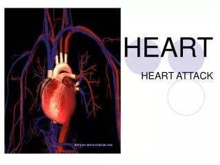
Heart
E N D
Presentation Transcript
CIRCULATORY SYSTEMFUNCTIONS: • Transport (nutrients, wastes, oxygen, CO2, hormones) • Immunity (leukocytes, antibodies) • Temperature regulation (when you are cold, blood vessels constrict; when hot, they dilate) • Penile erection
COMPONENTS OF CIRCULATORY SYSTEM • Heart • Blood • Blood vessels (arteries, capillaries, veins) • Lymph vessels and nodes
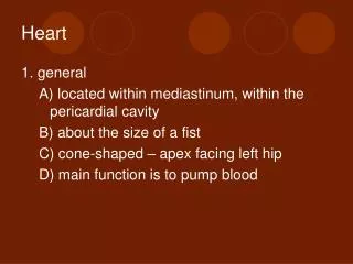
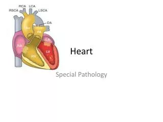
THE HEART • The heart is the simplest organ in the body. It does only one thing: pumps blood. • It beats 42 million times a year. • It’s about the size of your clenched fist. Some of you have big fists, some have smaller fists. • Its location is deep to the sternum. Take your left fist and place it on the sternum, then angle the bottom of your wrist to the left. When you say the Pledge of Allegiance, your hand is not over your heart. It’s not on the left, it’s in the center.
Location of the Heart in the Thorax Figure 18.2
Layers of tissues around the heart: Pericardium Heart muscle
PERICARDIUM Surrounds the heart (like a heart in baggie). The function is to lubricate the heart, so as it beats, it won’t rub against anything. • The pericardium is divided into two layers with a space between them filled with fluid: • PARIETAL PERICARDIUM • PERICARDIAL CAVITY • VISCERAL PERICARDIUM
PARIETAL PERICARDIUM This is actually the outermost layer of thoracic cavity. Two layers: • SEROUS LAYER (simple squamous epithelium). Watery fluid. • FIBROUS LAYER (moderately dense fibrous connective tissue)
VISCERAL PERICARDIUM (aka EPICARDIUM) Outermost layer of heart. It also has two layers: • SEROUS LAYER • FIBROUS LAYER
Layers of tissues around the heart: Pericardium Heart muscle
Structure of the Heart – Coverings Figure 18.3
MYOCARDIUM The heart muscle itself (myocardium) is made of what tissue? Cardiac muscle.
ENDOCARDIUM • The lining on the inside of the heart. Has two layers: • ENDOTHELIUM (simple squamous epithelium that provides a smooth surface for the blood to pass by) • Loose fibrous connective tissue (deep to the endothelium)
PERICARDITIS • Inflamed outer layer of heart. • Fluid accumulates in pericardial cavity, putting pressure on heart improper beat • Pericarditis can be caused by damage to the blood vessels blood leaks into pericardial cavity pressure improper beat. • Pericarditis can lead to pericardial friction rub, adhesions, and additional excess fluid in the pericardial cavity.
PERICARDITIS • CARDIAC TAMPONADE: In severe cases of pericarditis, or if there is a stab wound to the heart wall that causes fluid to exude into the pericardial cavity. • The excess fluid compresses the heart and diminishes the heart’s ability to pump. • Treatment is to stick a needle in the cavity and drain the fluid.
ENDOCARDITIS • More serious: • Bacteria enter bloodstream (dental procedures, IV drug abuse, catheter) damage to lining and valves blood clots. • Those who already have damaged heart valves need prophylactic antibiotics.
THE HEART IS TWO PUMPS: LEFT AND RIGHT LEFT PUMP: From lungs to body RIGHT PUMP: From body to lungs
The Pulmonary and Systemic Circuits Figure 18.1
Heart Chambers • Each pump has two types of chambers: ATRIUM and VENTRICLE Pulmonary artery SVC Pulmonary vein RA LA IVC LV RV Bicuspid valve Aorta Tricuspid valve
Blood Flow • Deoxygenated blood from body enters the RA through the superior and inferior vena cava. • It pours through the TRICUSPID (RIGHT AV) VALVE into the right ventricle. • Right atrium contracts, pushes blood into the right ventricle ventricle expands, then contracts with force. • To prevent the blood from going back up into the atrium, need a valve.
VALVES • Valves are like a swinging door that can only open one direction. But you can push against this door, since it’s only tissue. • But if you tie a rope to the doorknob, it won’t be able to go the wrong way. Rope = CHORDAE TENDONAE, which is attached to pieces of myocardium called PAPILLARY MUSCLES. • The contraction pulls on the chordae tendonae to close the valves, preventing a PROLAPSED VALVE (turned inside out).
Valves Figure 18.9a
Function of the Atrioventricular Valves Figure 18.9b
Heart Valves – Valve Structure Figure 18.8a
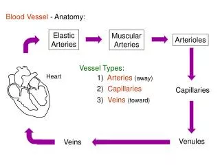
What is an artery? • An artery is a vessel that carries blood AWAY from the heart. It does not matter if it is oxygenated or deoxygenated blood. • A vein is a vessel that carries blood TOWARD the heart. It does not matter if it is oxygenated or deoxygenated blood.
Blood Flow • With the ventricular contraction, blood can go only one way: into the PULMONARY ARTERY (one of the few arteries with deoxy blood). Pulmonary artery SVC Pulmonary vein RA LA IVC LV RV Bicuspid valve Aorta Tricuspid valve
When the ventricles relax, the PULMONARY SEMILUNAR VALVE close to prevent blood from going from the pulmonary artery back into the right ventricle. . Pulmonary artery SVC Pulmonary vein Pulmonary semilunar valve RA LA IVC LV RV Bicuspid valve Aorta Tricuspid valve
Function of the Semilunar Valves Figure 18.10a, b
Blood Flow • When the ventricles relax, the PULMONARY SEMILUNAR VALVE closes to prevent blood from going from the pulmonary artery back into the right ventricle. • Do the semilunar valves have a chordae tendonae? • No; the blood is not being forced back (with a contraction), it just falls back with gravity, so there’s not as much pressure.
Blood then goes into lungs, gets oxygenated, and returns on the left side through the PULMONARY VEINS (one of the few veins with oxy blood), into the LEFT ATRIUM. Lungs Pulmonary artery SVC Pulmonary vein Pulmonary semilunar valve RA LA IVC LV RV Bicuspid valve Aorta Tricuspid valve
From the LEFT ATRIUM, it goes through the MITRAL VALVE (BICUSPID VALVE) into the LEFT VENTRICLE(there are also chordae tendonae here), which contracts. Pulmonary artery SVC Pulmonary vein Pulmonary semilunar valve RA LA IVC LV RV Bicuspid valve (Mitral) Aorta Tricuspid valve
Therefore, the left ventricle is the chamber which is responsible for generating the largest pressure upon contraction. Pulmonary artery SVC Pulmonary vein Pulmonary semilunar valve RA LA IVC LV RV Bicuspid valve (Mitral) Aorta Tricuspid valve
The blood then goes past the AORTIC SEMILUNAR VALVE, into the AORTA, and back to the body. The semilunar valves are located between the ventricles and the great arteries. Pulmonary artery SVC Pulmonary vein Pulmonary semilunar valve RA LA IVC LV RV Bicuspid valve (Mitral) Tricuspid valve Aorta Body Aortic semilunar valve
VALVES • CHORDAE TENDONAE • PAPILLARY MUSCLES • TRICUSPID VALVE • MITRAL VALVE (BICUSPID VALVE) • PULMONARY SEMILUNAR VALVE • AORTIC SEMILUNAR VALVE
SUMMARY OF BLOOD FLOW • Deoxy blood sup/inf vena cava R atrium tricuspid valve R ventricle pulmonary semilunar valve pulmonary artery lungs pulmonary veins Left atrium mitral (bicuspid) valve Left ventricle aortic semilunar valve aorta rest of body.
Heart Chambers Figure 18.5b
Heart Chambers Figure 18.5e
Inferior View of the Heart Figure 18.5d
Atrial septum Right atrium Left atrium Left ventricle Right ventricle Apex Interventricular septum
Aortic semilunar valve Mitral (bicuspid) valve Papillary muscles Cordae tendonae
Mitral Valve Stenosis • If there is stenosis (blockage) of the mitral valve, where will the blood back up into? • Answer: the pulmonary circulation. Lungs Pulmonary artery SVC Pulmonary vein Pulmonary semilunar valve RA LA IVC LV RV Bicuspid valve (Mitral) Tricuspid valve Aorta Body Aortic semilunar valve
HEART BEATS • The pressure of blood against blood vessel walls is called blood pressure. • Blood pressure is recorded systole over diastole. Normal resting blood pressure is said to be 120/80. When blood pressure is too high, it is called HYPERTENSION. • The sound your heart makes when it is beating is the sound of the blood hitting the valves after they are closed. • The heart normally beats at a rate of 60-80 beats per minute. A faster or slower heart rate is an indication of a problem.
HEART BEATS • The left and right ventricles contract at the same time = SYSTOLE. • When the ventricles are relaxed = DIASTOLE. • At which stage do the atria contract? Diastole.
HEART BEATS SYSTOLE: Ventricles contract Atria relax DIASTOLE: Ventricles relax Atria contract
HEART BEATS • Start of Systole: Closing of valves (tricuspid and mitral) causes blood to hit the valves, making a sound. Systole of the ventricle means that this chamber is contracting. • End of Systole: Closing of semilunar aortic and pulmonary valves causes blood to hit the valves, making a sound
HEART BEATS • Lub-Dub is the sound of the blood hitting the closed valves. • Start of Systole: Closing of the large valves (tricuspid and mitral) = “LUB” sound from blood hitting them. • End of Systole: Closing of semilunar valves (aortic and pulmonary)= “DUB” sound (“Dub”) from blood hitting them.
Heart Sounds Figure 18.11
Valve Problems • HEART MURMUR • If the valve leaks, it doesn’t close all the way • “Lub-squirt” • Most murmurs are benign; fairly common, esp. in babies and some adults.
Valve Problems • PROLAPSED VALVE is more serious. • Mitral valve is most likely to prolapse because it pumped the hardest. See how much thicker the left ventricle is? Mitral Valve Prolapse is the most common heart valve disorder. Might need artificial valve.