Download
1 / 33
350 likes | 560 Vues
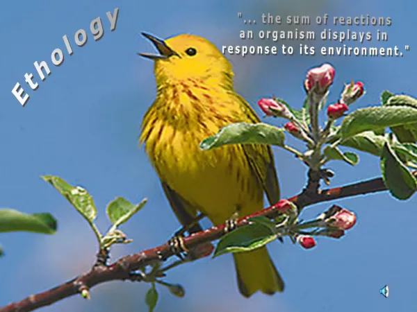
Biology 484 – Ethology Chapter 4a – Neural Mechanisms Controlling Behavior. Chapter 4 Woodhouse’s toad. What guides the behavior of these toads in mating? The Nervous System. 4.1 A complex response to simple stimuli.

E N D
Biology 484 – Ethology Chapter 4a – Neural Mechanisms Controlling Behavior
Chapter 4 Woodhouse’s toad What guides the behavior of these toads in mating? The Nervous System
4.1 A complex response to simple stimuli The male in “B” is attempting to mate with the thumb of the author of our book. The releaser of the behaviors for mating appear in this species to be a result of tactile stimulation on the undersurface of the insect. The shape of the female is roughly the same as the shape of the person’s thumb.
4.2 A simple rule of thumb governs this beetle’s mating behavior Colletes hederae displaying mating frenzy. The male can develop this “mating frenzy” under a wide array of conditions, all related to stimulation of the undersurface of the body. Here see a cluster of blister beetle larvae which the bee will also attempt to mate with, with surprising results.
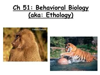
4.3 Pioneers in the study of animal behavior Tinbergen Lorenz von Frisch
4.4 Begging behavior by a gull chick The gull chick can elicit food regurgitation in the parent by tapping on the parent’s beak. The tapping behavior by the chick is neurally controlled as is the sensory detection of the tapping by the parent.
4.5 Effectiveness of different visual stimuli in triggering the begging behavior of herring gull chicks In this graph, we can see the components examined thought to be responsible for the elicitation of the pecking behavior in the chick. Note the numbers are relative percentages in each example.
4.6 Instinct theory Tinbergen originated the INSTINCT THEORY along with Lorenz. The basics of the theory are that simple stimuli (such as the red dot) can “release” a complex behavior in another bird such as the chick’s tapping behavior (begging behavior).
4.7 A code breaker The cuckoo in this image has been able to figure out the necessary behavioral pattern to guide the parent bird ( a reed warbler ) to give it food. In effect, the cuckoo has learned to be a behavioral code breaker.
The male Australian Beetle will attempt to mate with virtually anything that is of a similar color to itself. On the left is a beer bottle, on the right a road sign. Thought Question: This behavior seems to be not appropriate, how/why would you hypothesize the behavior remains in the species?
Santiago Ramon Y. Cajal (1852-1934) Founding Scientist in the Modern Approach to Neuroscience. Received Nobel Prize in 1906
Figure 11.3: Neuroglia, p. 390. Capillary Neuron (b) Microglial cell (a) Astrocyte Nerve fibers Myelin sheath Fluid-filled cavity Process of oligodendrocyte Brain or spinal cord tissue (c) Ependymal cells Cell body of neuron (d) Oligodendrocyte Satellite cells Schwann cells (forming myelin sheath) Nerve fiber (e) Sensory neuron with Schwann cells and satellite cells
Figure 11.5: Relationship of Schwann cells to axons in the PNS, p. 394. Schwann cell cytoplasm Schwann cell plasma membrane Axon Myelin sheath Schwann cell nucleus (a) Schwann cell cytoplasm Axon Neurilemma (b) (d) Neurilemma Myelin sheath (c)
Neurotransmitter chemical attached to receptor Receptor Na+ Na+ Chemical binds K+ K+ Closed Open (a) Chemically gated ion channel Na+ Na+ Membrane voltage changes Closed Open (b) Voltage-gated ion channel
Figure 11.7: Measuring membrane potential in neurons, p. 399. Voltmeter Plasma membrane Ground electrode outside cell Microelectrode inside cell Axon Neuron
Figure 11.8: The basis of the resting membrane potential, p. 399. Cell exterior Na+ Na+ 15 mM Cell interior Na+ Na+ ion K+ 150 mM Diffusion Na+–K+ pump us Diff Cl– 10 mM -70 mV Na+ Na+ A– 100 mM Na+ 150 mM K+ Na+ Plasma membrane A– 0.2 mM Na+ K+ K+ 5 mM Cl– 120 mM K+ Cell interior Cell exterior K+ K+
Figure 11.9: Depolarization and hyperpolarization of the membrane, p. 400. Depolarizing stimulus Hyperpolarizing stimulus +50 +50 Inside positive 0 0 Inside negative Depolarization Membrane potential (voltage, mV) Membrane potential (voltage, mV) –50 –50 Resting potential –70 –70 Resting potential Hyper- polarization –100 –100 0 1 2 3 4 5 6 7 0 1 2 3 4 5 6 7 Time (ms) Time (ms) (a) (b)
Figure 11.10: The mechanism of a graded potential, p. 401. Depolarized region Stimulus Plasma membrane (a) Depolarization (b) Spread of depolarization
Figure 11.11: Changes in membrane potential produced by a depolarizing graded potential, p. 402. Active area (site of initial depolarization) Membrane potential (mV) – 70 Resting potential Distance (a few mm)
Figure 11.12: Phases of the action potential and the role of voltage-gated ion channels, p. 403. Outside cell Outside cell Na+ Na+ Inside cell K+ Inside cell K+ Repolarizing phase: Na+ channels inactivating, K+ channels open Depolarizing phase: Na+ channels open 2 Action potential +30 3 0 Relative membrane permeability 2 PNa Membrane potential (mV) PK Threshold –55 1 1 4 –70 0 1 2 3 4 Time (ms) Outside cell Sodium channel Potassium channel Outside cell Na+ Na+ Activation gates K+ Inside cell K+ Inside cell Inactivation gate Hyperpolarization: K+ channels remain open; Na+ channels resetting 4 1 Resting state: All gated Na+ and K+ channels closed (Na+ activation gates closed; inactivation gates open)
Figure 11.13: Propagation of an action potential (AP), p. 405. Voltage at 2 ms +30 Membrane potential (mV)) Voltage at 0 ms Voltage at 4 ms –70 (a) Time = 0 ms (b) Time = 2 ms (c) Time = 4 ms Resting potential Peak of action potential Hyperpolarization
Figure 11.14: Relationship between stimulus strength and action potential frequency, p. 406. Action potentials +30 Membrane potential (mV) – 70 Stimulus amplitude Threshold Voltage 0 Time (ms)
Figure 11.15: Refractory periods in an AP, p. 406. Absolute refractory period Relative refractory period Depolarization (Na+ enters) +30 0 Repolarization (K+ leaves) Membrane potential (mV) After-hyperpolarization –70 Stimulus 0 1 2 3 4 5 Time (ms)
Figure 11.16: Saltatory conduction in a myelinated axon, p. 407. Node of Ranvier Cell body Myelin sheath Distal axon
Figure 11.17: Synapses, p. 409. Cell body Dendrites Axodendritic synapses Axosomatic synapses Axoaxonic synapses Axon (a) Axon Axosomatic synapses Soma of postsynaptic neuron (b)
Figure 11.18: Events at a chemical synapse in response to depolarization, p. 410. Neurotransmitter Na+ Ca2+ Axon terminal of presynaptic neuron Action Potential Receptor 1 Postsynaptic membrane Mitochondrion Postsynaptic membrane Axon of presynaptic neuron Ion channel open Synaptic vesicles containing neurotransmitter molecules 5 Degraded neurotransmitter Na+ 2 Synaptic cleft 3 4 Ion channel closed Ion channel (closed) Ion channel (open)
4.10 The eyestalks of a fiddler crab point straight up The eyestalks in this crab point upwards and determine its field of view. The stalks will change the perspective it views compared to other many other more standard positions. Question to Ponder…. What can you hypothesize about the role/benefit for this placement for the crab compared to other eye positions?