Class : Arachnida Order : Acarina Mites
511 likes | 2.54k Vues
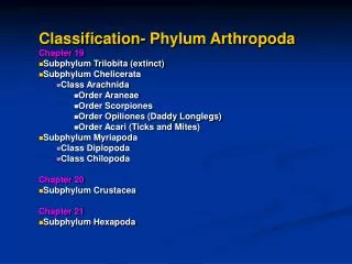
Class : Arachnida Order : Acarina Mites. By Assist. Lecturer Maytham A. Alwan. Phylum: Arthropoda. phylum. Arthropoda. Class. Arachnida. Insecta. Order. Others e.g. Hemiptera. Acarina. Diptera. Phthiraptera. Siphonaptera. Ticks. Mites. flies. lice. fleas. bed bugs.

Class : Arachnida Order : Acarina Mites
E N D
Presentation Transcript
Class : ArachnidaOrder : AcarinaMites By Assist. Lecturer Maytham A. Alwan
Phylum: Arthropoda phylum Arthropoda Class Arachnida Insecta Order Others e.g. Hemiptera Acarina Diptera Phthiraptera Siphonaptera Ticks Mites flies lice fleas bed bugs
Acarina : Mites • The parasitic mites are small, most being less than 0.5 mm long, though a few blood-sucking species may attain several mm when fully engorged. causing various forms of the condition generally known as mange. • Although, like the ticks, mites are obligate parasites, they differ from them in the important respect that most species spend their entire life cycles, from egg to adult, on the host so that transmission is mainly by contact. • The mites have a complex taxonomy, occupying at least eight different families, and for veterinarians. Mites can be divided into two major groups: sarcoptiform and nonsarcoptiform mites. Nonsarcoptiform mites includes demodecidae (Demodex canis) • The sarcoptiform mites can be subdivided according to their location on the host as burrowing(Tunneling)
Mites Burrowing Mites The three important burrowing genera, Sarcoptes, Notoedres and Knemidocoptes, belong to a single family, the Sarcoptidae Non-burrowing (surface dwelling) mites (Psoroptes, Chorioptes, Otodectes), belong to a family, the Psoroptidae classification Order : Acarina Family: Sarcoptidae Genus: Sarcoptes
Sarcoptes • Sarcoptesis well known in both human and veterinary medicine as a cause of mange, the disease in man being generally known as scabies. • Hosts: • All domestic mammals and man. • Species: • S. scabieivarhominis(human), S. scabieivar. canis( dogs), it can also infest other mammals, including cats, pigs, foxes, rabbits and guinea pigs for varying periods of time. S. scabieivar. suis( pigs), S. scabieivarbovis( cattle), S. scabieivar. equi( horses),S. scabieivarovis( sheep),S. scabieivarcaprae( goats). • Distribution: Worldwide.
Sarcoptes Morphology Adult scabies mites are spherical(rounded) , dorsoventrally flattened up to 0.4 mm in diameter, and the cuticle is striated bearing a central patch of raised scales with four pairs of short legs having suckers on a long unjointed stalk on pairs 1st and 2nd in the case of female and on pairs 1st, 2nd and 4th in male. The remaining legs all terminate in long, hair-like setae.
Sarcoptes life cycle Sarcoptesscabieimites complete their entire four-stage life cycle : egg, larvae, nymph and adult on the host, and do not survive for long periods in the environment. Male and female mites breed on the skin surface. Males do not burrow into skin. The females penetrate the keratinized layers of the skin and burrow tunnels through the epidermis over a 10-15 day period. The female deposits 40 to 50 eggs within the tunnel. After egg deposition, the female dies. Six-legged larvae emerge from the eggs in (3-10) days and exit the tunnel to wander on the skin surface. These larvae molt to the eight-legged nymphal stage within tiny pockets called molting pouches in the epidermis. Nymphs become sexually active adults in 12 to 17 days and the life cycle begins again.
Sarcoptes Clinical Signs In sheep, Sarcoptesscabiei variety ovis affect the non-woolly skin, and the lesions typically start on the lips or nostrils. The lesion on the nostrils also extends around the eyes, the supraorbitalfossae and in some cases over entire face and head. The clinical signs can be summarized in itch , intense pruriticerythematous dermatitis and papular rash as the alopecia spreads. The rash often becomes generalized leading to hair loss due to which animals loose much of the grazing time and hence loose general body condition (weight loss), secondary bacterial infection where severe untreated cases can be fatal , death is usually the result of secondary bacterial infections.
Sarcoptes • Pathogenesis SarcoptesfemaleMites burrow into the epidermis and feed on tissue fluids. The burrowing and feeding of the mite cause irritation and consequential scratching, leading to inflammation and exudation to form crusts. If left untreated, the skin wrinkles and thickens with proliferation of the connective tissue followed by depilation. Death of the animals may occur in severe mite infestation.
Non burrowing mites Including: psoroptes, chorioptes, otodectes Family : Psoroptidae Genus : Psoroptes. Species : P. communisvarovis.(sheep) P. communisvarcaprae (goat) P. equi (equine) P. natalensis ( cattle and horses) P. cuniculi (ear mite of rabbits, goats, horses and sheep)
Psoroptes • Mites of the genus Psoroptes are obligate, non-burrowing, astigmatid ectoparasites of mammals, of particular economic importance in domestic animals where they cause clinical psoroptic mange. • Morphology • Mature female Psoroptes up to 0.75 mm, with a striate cuticle. A noticeable anterodorsal cuticular plate is present behind the mouthparts, and the midventral ovipore is an inverted U-shape. Males are about one-fourth smaller, and they have a pair of posteroventral adanal suckers, and two terminal posterior lobes, each equipped with four setae of varying lengths and structures. Nymphs and larvae are somewhat similar to adults but progressively smaller, and all Psoroptes are pearly white in color.
In all stages, the anterior two pairs of legs are thicker and more robust than the posterior pairs, which are thinner, and in the male, shortened in the fourth pair. Legs I and II terminate in pretarsal empodial suckers on long, segmented pedicels in both sexes, with similar structures on legs IV of the female and legs III of the males. The female’s third tarsus ends in two long, whip-like setae, and the male has a single short seta on tarsus IV. Male of psoroptes female of psoroptes
Psoroptes life cycle The life cycle begins with the deposition of the eggs on the skin at the margin of the lesion. These hatch in 1 to 5 days producing 6-legged larvae. The larvae feed for 2 to 3 days and then molt and become 8-legged nymphs. The nymphal stage lasts 3 to 4 days when they molt and become mature males and females which mate immediately. The fertilized female molts and then lays eggs. The cycle from egg to adult is compeleted in 12 days. Females may live as long as 42 days on sheep.
Psoroptes Clinical signs Psoroptes mites usually affect the shoulders, back, sides, lower cervical and upper thoracic region. Sheep scab is characterised by intense itching and pruritus. The flock may be seen repeatedly rubbing their shoulders and flanks along the ground or against fences, foot stamping, clawing at their flanks, or biting their shoulders and sides. Early cases of sheep scab are characterised by red sores with serous exudation, yellow/orange staining wool fibres and displacement of small tufts of wool. As the disease progresses, the serous exudation increases in severity and extent, extending over the shoulders to the chest wall, back and flanks.
Diagnosis 1. Clinical manifestations They are summarized by hair loss, crusty or scaly skin lesions, dermatitis, thickened skin, blisters, scurf, and pruritus. 2. Laboratory tests : which include two methods a. Direct method: the skin scraping place in a test tube and add 5-10 ml of KOH 10%. The tubes are placed in a water bath with 60-80˚c for 15 minutes then transported to a centrifuge in a speed of 1500-2000 rounds per minute for 5 minutes then the supernant is discarded by an automatic pipette and the sediment is mixed well in a test tube. Then some drops are drawn from the sediment with a pipette and placed on a glass slide covered with a cover slide and examined under microscope with power 10 x, 40 x, 100 x to confirm the presence of parasite. b. Indirect method: 1. ELISA 2. Polymerase Chain Reaction (PCR)
Treatment • in animals mite infestations are treated with • 1.acaricides including lime sulfur, the animal may be bathed first with an antiseborrheic shampoo to remove crusts and debris. • 2.Ivermectin is used to treat some mites, and doramectin has been used for sarcoptic mange. Selamectin has recently been reported to be effective for sarcoptic mange. If the mites can survive for more than a few days in the environment, the animal’s surroundings must also be treated with an insecticide or acaricide .
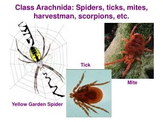
Mites some differences between Ticks & Mites Ticks Large in size, can be seen by the nakedEyes All of them parasitic Covered by scutum Armed hypostome Parts of it's lifecycle is free living Lay it's egg on the ground in cracks or under the rocks Body is not covered by hair Microscopic in size Some of them free living & other parasitic on plants Scutum not present and the skin is leathery not armed hypostome All it's lifecycle on the body of the host Lay it's egg in tunnels at the site of infection on the body of the host Body is covered by hair