Download
1 / 92
1.03k likes | 1.35k Vues
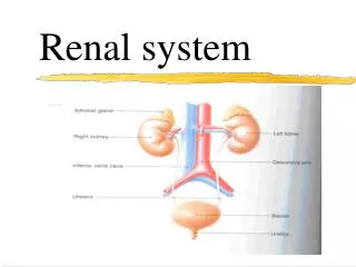
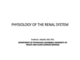
RENAL SYSTEM. Organs of the Renal System. Kidneys Ureters Urinary bladder Urethra Not many structures, but very important!. Figure 23.1a. Functions of Urinary System. Regulate electrolytes (K+, Na+, etc ) Regulate pH in blood Regulate blood pressure

E N D
RENAL SYSTEM mehdi jalali majd
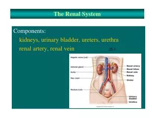
Organs of the Renal System • Kidneys • Ureters • Urinary bladder • Urethra • Not many structures, but very important! mehdi jalali majd Figure 23.1a
Functions of Urinary System • Regulate electrolytes (K+, Na+, etc) • Regulate pH in blood • Regulate blood pressure • Regulate blood volume (removes excess fluid) • Removing metabolic wastes • Urea, uric acid, and creatinine • This is the least important of the kidney’s functions. You can survive for a few weeks without excreting waste products in the urine, but hour by hour, the other functions are more important. mehdi jalali majd
Relationship of the Kidneys to Vertebra and Ribs They are retroperitoneal and are located in the abdominal cavity. They are at the level of T12 to L3, so they are at the costal margin, and the floating ribs protect them a little. Even though they are protected by thoracic ribs, they are NOT in the thoracic cavity because they are below the diaphragm. mehdi jalali majd Figure 23.1b
Position of the Kidneys with in the Posterior Abdominal Wall mehdi jalali majd Figure 23.2a
STRUCTURES WITHIN THE KIDNEY • The RENAL CAPSULE surrounds the kidney, made of dense fibrous connective tissue. • A layer of adipose tissue surrounds the capsule, called PARARENAL FAT (ADIPOSE). It cushions and protects. • Around that is a connective tissue layer called the RENAL FASCIA, made of loose connective tissue. It anchors the kidney to the surrounding peritoneum and abdominal wall. It is not very strong; jumping up and down can cause tearing. mehdi jalali majd
Vibration Platform Machine:No longer used in the USA because it damages kidneys! mehdi jalali majd
These are still around in gyms in other countries…beware! mehdi jalali majd
Gross Anatomy of the Kidneys • Renal cortex (most superficial layer) • Renal medulla • Renal pyramids (drain into the calyx) • Renal pelvis • Calyx (drains into hylus ureter) • Ureter mehdi jalali majd
Renal fascia Interlobar arteries Interlobular arteries Arcuate arteries mehdi jalali majd
Internal Anatomy of the Kidneys Interlobar artery mehdi jalali majd Figure 23.3b
Blood Supply to Kidney AORTA RENAL ARTERY SEGMENTAL ARTERIES INTERLOBAR ARTERIES ARCUATE ARTERIES (form arcs) INTERLOBULAR ARTERIES INTERLOBULAR VEIN ARCUATE VEIN INTERLOBAR VEINS SEGMENTAL VEINS RENAL VEIN INF. VENA CAVA mehdi jalali majd
Internal Anatomy of the Kidneys Interlobar artery mehdi jalali majd Figure 23.3b
Microscopic Anatomy of the Kidneys • Just like the functional unit of the lungs is the alveolus and the functional unit of the liver is the lobule, the functional unit of the kidney is the NEPHRON. • Each kidney has about 1 million nephrons. • Each one carries out all of the various functions of the kidneys. mehdi jalali majd
Microscopic Anatomy of the Nephron • GLOMERULUS WITH A CAPSULE • PROXIMAL CONVOLUTED TUBULE • LOOP OF HENLE • DESCENDING LIMB • Thick portion • Thin portion • ASCENDING LIMB • Thick portion • Thin portion • DISTAL CONVOLUTED TUBULE • COLLECTING DUCT mehdi jalali majd
Nephron mehdi jalali majd
Position of Nephron in Kidney mehdi jalali majd Figure 23.4a
Glomerulus of a Nephron mehdi jalali majd
Juxtaglomerular Apparatus mehdi jalali majd • The distal convoluted tubule passes next to the glomerulus to form the juxtaglomerular apparatus (juxta means “next to”). • The juxtaglomerular apparatus (JGA) consists of cells located in and around the glomerulus and the glomerular capsule.
Juxtaglomerular Apparatus mehdi jalali majd • If blood pressure is too low, the JGA releases adenosine, which causes vasoconstriction of the afferent arteriole. This will slow the filtration rate so less water is lost, and blood pressure increases.
Juxtaglomerular Apparatus: Juxtaglomerular Cells mehdi jalali majd • if the blood pressure is still too low after adenosine has caused vasoconstriction, the JGA secretes the hormone renin. • Renin causes more sodium to be reabsorbed, and water follows, so blood volume increases, so blood pressure increases.
GLOMERULUS • The glomerulus is the first part of the nephron, where the filtration occurs. That means plasma leaks out of the capillaries and into the convoluted tubules. • The glomeruli are located only in the renal cortex. • A glomerulus (“ball of yarn”) is a tuft of capillaries surrounded by a glomerular capsule (Bowman’s capsule) made of simple squamous epithelium. The capillaries fits in the capsule like a fist punched into an underinflated balloon. • The capsule collects the plasma from the capillaries and drains it into the convoluted tubules, which empty into a collecting duct, which exits the body. • The plasma is further filtered along the way. The good nutrients are reabsorbed back into the blood. mehdi jalali majd
Glomerulus of a Nephron mehdi jalali majd
Normally at the end of the capillary bed you have venuoles. But this is the only part of the body that is different: here we have another arteriole, called the EFFERENT ARTERIOLE, which takes blood away from the glomerulus. • The efferent arteriole drops down straight, next to the Loop of Henle. While it is straight, it is called VASA RECTA (straight capillaries). • There are capillaries that come off the vasa recta which surround the loop of Henle. Here, they are called peritubular capillaries. They then leave the area to become the interlobular vein, which leaves the kidney. mehdi jalali majd
Nephron mehdi jalali majd
Distal convoluted tubule Efferent arteriole Afferent arteriole Bowman’s capsule Proximal convoluted tubule mehdi jalali majd
FUNCTION OF THE NEPHRON • Blood comes in from the AFFERENT ARTERIOLES. • Plasma leaks out and enters the glomerular capsule. The plasma contains nutrients, which need to be reabsorbed, as well as waste products. • As the plasma moves through the proximal convoluted tubule, all of the nutrients, and most of the water, and most of the ions are reabsorbed back out of the nephrons and into the blood. • In the Loop of Henle, almost all of the rest of the water and salt are reabsorbed into blood. • Everything that is not reabsorbed (waste products) goes into the collecting duct and is excreted as urine.This is also how the water-salt balance is maintained, as well as the acid-base balance. The kidneys can remove or retain acids as well. mehdi jalali majd
Nephron mehdi jalali majd
FUNCTION OF THE NEPHRON • In the distal convoluted tubule, the rest of the water and salt are removed. • The rest of the liquid goes into the collecting duct. • The distal convoluted tubule and the collecting duct fine-tune the water and salt absorption and excretion. If you are well hydrated, the water will be allowed to leave as urine. • If you are thirsty, the water will be absorbed. The purpose of the peritubular capillary bed is to absorb these things from the nephron tubules and put them back into the blood. mehdi jalali majd
mehdi jalali majd Figure 23.5
Renal Corpuscle and the Filtration Membrane mehdi jalali majd Figure 23.6c
Function of the Nephron mehdi jalali majd NEPHRON VIDEO http://www.youtube.com/watch?v=aQZaNXNroVY 6 Tips for healthy urinary system: http://urology.about.com/od/infections/tp/5tipsforurinehealth.htm Lupus Nephritis
Diuretics • Diuretics are medicines that increase the amount of urine that is produced. • People who have high blood pressure might be prescribed diuretics to decrease the blood volume. • Alcohol is a diuretic and this is what contributes to the symptoms of a hangover. The best way to prevent a hangover after drinking is to consume a lot of water before you go to bed. • Caffeine is also a diuretic, so coffee, energy drinks, and regular Coca-cola are diuretics. • You should drink one cup of water for every cup of those beverages to prevent dehydration. mehdi jalali majd
HISTOLOGY OF THE NEPHRON • PROXIMAL CONVOLUTED TUBULES • This area absorbs all nutrients and 2/3 of the water and salt. Only about 1% of the fluid filtered by the kidney actually becomes urine. • LOOP OF HENLE • This is where 1/3 of water and salt is reabsorbed. It is located in the renal medulla. mehdi jalali majd
Urine Production • Filtration – filtrate of blood leaves kidney capillaries • Reabsorption –nutrients, water, and essential ions reclaimed • Secretion – active process of removing undesirable molecules mehdi jalali majd
mehdi jalali majd Figure 23.9a
Collecting Duct (tubule) • Receives urine from distal convoluted tubules, empties into the calyx, which empties into the ureter. mehdi jalali majd
Ureters • These are long tubes that connect the renal pelvis to the urinary bladder • MUCOSA • TRANSITIONAL EPITHELIUM (for expansion) • LAMINA PROPRIA (has elastic tissue to recoil) • MUSCULAR LAYER (smooth muscle) • INNER CIRCULAR • OUTER LONGITUDINAL • ADVENTITIA • Loose connective tissue • Provides protection, strength for organs, and attaches ureters to surrounding structures mehdi jalali majd
URINARY BLADDER • The histology of the urinary bladder is identical to the ureter except the mucosal layer has folds called RUGAE, which allow for expansion. • The rugae have the same basic function as transitional epithelium—accommodating stretch as the bladder fills. • You can hold up to one liter of urine, although at 500 ml, you’ll be dancing. mehdi jalali majd
Urinary Bladder • Rugae: allows for expansion • -A full bladder is roughly the size of a soft ball mehdi jalali majd Figure 23.14
URINARY BLADDER • The muscle layer that makes up the urinary bladder is thicker than the ureters, and is called the DETRUSOR MUSCLE, which contracts to allow emptying of the urinary bladder. • The function of the urinary bladder is just to store urine. mehdi jalali majd
Detrusor Muscle mehdi jalali majd
URETER entrance to BLADDER • The URETERS enter in at the base of the urinary bladder, not the top. • As the bladder fills, the urine presses down on the ureters to prevent urine from backing up into the kidneys. • Da Vinci surgery • http://www.davincisurgery.com/da-vinci-urology/da_vinci_partial_nephrectomy.php mehdi jalali majd
BLADDER • The TRIGONE is a triangular area where the ureters come in and the urethra goes out. • Between the urethra and the urinary bladder are two sphincters: • INTERNAL URETHRAL SPHINCTER: smooth muscle • EXTERNAL URETHRAL SPHINCTER: skeletal muscle. Although it is primarily under voluntary control, it will release if the urine volume is too much. • If the patient is in a coma or under anesthesia for a long time, the internal sphincter will be closed, like when you are asleep, so a catheter is needed to open it to drain the urine out. mehdi jalali majd
Bladder Stones mehdi jalali majd
Pelvic Ultrasound mehdi jalali majd Attachment
Urethra • Drains the urine to the outside. • Its histology is the same as the ureter • Females: 4cm • Males 20 cm (varies with mood) • Therefore, women (esp. little girls) are more susceptible toUTI. mehdi jalali majd
Problems • URETHRITIS = infection and inflammation of the urethra • PYELITIS= infection of the renal calyxes • CYSTITIS = infection of the urinary bladder. • CHOLEOCYTITIS = infection of the gall bladder mehdi jalali majd
UREA • Urea is a waste product of amino acid metabolism. • Remember, proteins are made of amino acids, so when you break down proteins, you break them down into amino acids, and the waste product left over is urea. • This is the main waste product in urine. • Eating excessive proteins can cause kidney damage, since their breakdown products are acidic. mehdi jalali majd