Download
1 / 37
1.31k likes | 5.34k Vues
Hemacytometer and Manual Cell Counts. Kimberly Murray University of Phoenix EDTC 560: Applications of Multimedia and Web Page Design June 4, 2007. Quote of the Day.

E N D
Hemacytometer and Manual Cell Counts Kimberly Murray University of Phoenix EDTC 560: Applications of Multimedia and Web Page Design June 4, 2007
Quote of the Day • “Education is a technique employed to open minds so that they may go from profound ignorance to thoughtful uncertainty” (Mitner, 1999).
Objectives • Upon completion of this course, the student will: • Identify the dimensions of the Neubauer hemacytometer. • Identify sources of error in counting. • Calculate manual cell counts.
Hemacytometer Counting Chamber • A precision tool designed to standardize counting. • Used to count cells in peripheral blood or other body fluids.
Basic hemacytometer formula Number of cells counted X dilution factor total volume of the cells counted total volume = # primary squares x 0.1 (depth) This formula can be used with any counting chamber as long as one knows the dimensions for the chamber
Hemacytometer Components Cover slip –NBS standardized Moat Fill trough Chambers or Counting grid Diagrams by K. P. Murray, 2007
Sample is placed between cover slip and chamber The design of the hemocytometer is to have the support for the cover slip at a specific height, resulting in a known depth of exactly 0.1mm. Cover slip –NBS standardized Moat 0.1 mm Depth National bureau of standards must be met for both the chamber and cover slip. Sample Diagrams by K. P. Murray, 2007
Do Not Use Disposable Cover Slip Sample floats coverslip Do not use a regular cover slip especially for viscous fluids. The sample will float disposable coverslips and the depth will be greater than 0.1mm. Diagrams by K. P. Murray, 2007
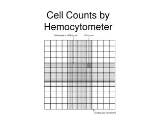
Neubauer Hemacytometer Dimensions • Each side (chamber) has a ruled area • 3 mm wide and 3 mm long • Divided into nine squares • The large squares are subdivided into 16 • The middle square is subdivide into 25 • Each of the 25 is again subdivided into 16 • When the dimensions are known, one will always be able to calculate the area counted.
Dimensions of the Neubauer Note: Center primary square has different divisions Primary squares 1.0 mm Secondary Squares 0.2mm 0.25mm Tertiary squares 0.0125 mm Diagrams by K. P. Murray, 2007
Hemacytometer Quality Control (QC) • Ensures accuracy • Performed by duplicating the counting area exactly. • For example: • If squares 1,3,5,7,9 are counted. • Then 10,12,14,16,18 are counted on the other side. Diagrams by K. P. Murray, 2007
Area Counted Depend on Cell Type WBC WBC . . Platelet RBC RBC RBC RBC RBC . . WBC WBC Diagrams by K. P. Murray, 2007
RBC Count Method • Count the five inner tertiary squares on both sides of the chamber • on Peripheral Blood • Check difference in counts • Calculate RBC count using the basic hemacytometer formula . . RBC RBC RBC RBC RBC . . Diagrams by K. P. Murray, 2007
RBC Count Method • If a Body Fluid count is less than 100 cells • count all 9 squares on both sides to provide greater accuracy. • Adjust the hemacytometer calculation accordingly . . . . Diagrams by K. P. Murray, 2007
WBC Count Method • Count the four outer secondary squares on both sides of the chamber • Check difference in counts • Calculate WBC count using the basic hemacytometer formula WBC WBC WBC WBC Diagrams by K. P. Murray, 2007
WBC Count Method • If WBC count is less than 1000 cells • count all 9 squares on both sides to provide greater accuracy. • Adjust the hemacytometer calculation accordingly Diagrams by K. P. Murray, 2007
Neubauer Hemacytometer Calculation Cells X dilution = cells/L squares X 0.1 Note: 0.1 mm depth is constant in Neubauer Hemacytometer
Unopette – used today for manual counts • Components • Reservoir • Capillary pipette • Protective sleeve that also is used to open the reservoir. • Pre-measured diluents in vials. • Capillary pipette is specific for the diluent used. • The capillary tubes can not be interchanged. Picture by K. P. Murray, 2007
RBC vial Isotonic or physiologic saline 1/200 dilution WBC only vials Turk’s solution 1/20 dilution WBC/PLT vials Ammonium oxalate --11.45 g Sorenson’s buffer--1.0g Thimerosal -- 0.1g (preservative) Distilled H2O qs to 1 liter 1/100 dilution Unopette System For RBC, WBC and PLT counts RBC can not resist osmotic pressures, as the WBC can, therefore RBC counts need isotonic solutions Manual RBC counts in PB rarely done Always performed on other body fluids
Unopettes Note: different final dilutions
Loading Chamber using a Unopette Picture by K. P. Murray, 2007
“Wet Box” prevents solution from drying out and altering count • Settle cells to the same optical plane • WBC • 2-3 minutes • PLT • 10 minutes (take the longest to settle) • RBC • 2-3 minutes Picture by K. P. Murray, 2007
Acceptable differences between chambers of the hemacytometer • No more than 10 cell variation between the 4 squares for the WBC count. • The difference between the highest and the lowest number should not be greater than 25 cell for the 10 tertiary squares (5 squares per side) the RBC count. Diagrams by K. P. Murray, 2007
How to Tell If Sample has Uneven Distribution • Difference between individual squares exceeds 10% • Difference between each chamber count exceeds 20 % • REPEAT COUNT with a freshly loaded hemacytometer, if the count does not match the above criteria.
WBC Count Reporting • Report WBC • Number (period) one decimal X 103/ L • Number. decimal X 103/ L • Example: 10.9 X 10³/ L WBC has one decimal place, if sample is peripheral blood. For other body fluids (BF), report whole number, no scientific notation or abbreviations.
RBC Count Reporting • Report RBC • Number (period) two decimals X 106/ L • Number. two decimals X 106/ L • Example: 4.13 X 10³/ L RBC has two decimal places, if peripheral blood. For other body fluids (BF), report whole number, no scientific notation or abbreviations.
Platelet Count Reporting • Report PLT • Whole number (no decimal) X 103/ L • Example: 428 X 103/ L Platelet count does not have a decimal point. Other body fluids do not report platelet counts.
Platelet Count Reporting • Verbal reporting always give the full number. • For example, “Doctor Jones, the platelet count on Ms. Smith is 428,000”. • DO NOT just say the count is 428. • The doctors or the nurse may think only four hundred (transfusion time!!!), rather than a normal count of 400 thousand.
Reference Units Notice the similarities and differences between units
Sources of Error • Failure to mix sample prior to dilution • Pipeting technique • Uneven distribution in chamber • Failure to remove excess blood from outside of pipette • Squeezing out some fluid from reservoir [Unopette]
Sources of Error • Cell aggregates (clots) • Bacteria ( may be mistaken for plt) • Dirt, talc, debris • Drying of the chamber • taking too long to count • Falsely increase counts
Fill problems Correct fill Coverslip Air bubbles Overfill Underfill Diagrams by K. P. Murray, 2007
Problem 1 • A blood sample was diluted1:20. • On a Neubauer hemacytometer • 140 WBC counted in the four outer squares on side A. • 147 WBC counted on the same four squares on side B. • What is the WBC count?
Problem 1 answer • WBC 140 and 147 ‘match’ and average 143.5 • Depth = 0.1 (always with a Neubauer) • 143.5 cells x 20 (dilution factor) 4 (squares) x 0.1(depth) WBC = 7,175 WBC =7.2 x 10 3 /dL
Problem #2 • A tech counted both side of the hemacytometer on an undiluted spinal fluid sample. • 22 WBC were counted and no RBC. • What is the WBC count? • How is it reported?
Problem #2 solution • Both side area = 18 mm2 {(1x1) (9+9)} • Depth = 0.1 • Both side count = 22 • Dilution = 1 • Answer: WBC = 12 cells / dL • NOTE: no decimal or scientific notation because it is a body fluid and one does not report a fraction of a cell.
References • Barry T. Mitzner, (1999), D.V.M, Southeast Vetlab, Inc., Retrieved on July 12, 2003 from http://www.spcollege.edu/hec/vettech/VTDE/Hematology%20methods%20for%20the%20offi/ppframe.htm • Murray, K.P.,(2007) Hematology Lab Photographs (personal collection) • Murray, K.P.,(2007) Hematology Diagrams and Animations (personal collection) END