Download

1 / 24
240 likes | 378 Vues
LOWER EXTREMITY X-RAYS, MRIS AND ANGIOGRAMS 2008. Sacroiliac Joint. Anterior superior iliac spine. Neck of Femur. Pubic Symphysis. Greater Trochanter. Inferior ramus of pubis. Ischial Tuberosity. Lesser Trochanter. 1 X-ray Pelvis and Proximal Femur (same X-ray as studied in

E N D
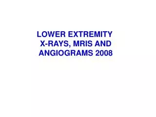
LOWER EXTREMITY X-RAYS, MRIS AND ANGIOGRAMS 2008
Sacroiliac Joint Anterior superior iliac spine Neck of Femur Pubic Symphysis Greater Trochanter Inferior ramus of pubis Ischial Tuberosity Lesser Trochanter
1 X-ray Pelvis and Proximal Femur (same X-ray as studied in Abdomen and Pelvis) 1. Anterior Superior Iliac Spine 2. Acetabular rim of pelvis 3. Sacral foramina for Spinal nerve 4. Pubic symphysis 5. Inferior Ramus of Pubis 6. Obturator Foramen 7. Ischium 8. Head of Femur 9. Neck of Femur 10. Greater Trochanter 11. Lesser Trochanter
#10 Venogram Iliac and Femoral Veins 1. External Iliac Vein 2. Internal Iliac Vein 3. Valve Cusps in Great Saphenous and Femoral Veins
#9 Arteriogram of Femoral Artery 1. Head of femur (note relationship to femoral artery #2) 2. Femoral artery 3. Femoral artery in thigh 4. Profunda Femoris Artery 5. Lateral Femoral Circumflex Artery 6. First perforating branch of profunda femoris artery
# 2 X-RAY OF KNEE JOINT, TIBIA AND FIBULA 1. Medial condyle of femur 2. Medial condyle of tibia 3. Intercondylar eminence of tibia 4. Lateral condyle of femur 5. Head of fibula
#3 Lateral X-ray of Knee Joint 1. Patella 2. Condyle of femur 3. Medial condyle of tibia 4. Head of fibula
#1 - Lateral X-ray of Knee with Fabella 1- Fabella - this is a sesamoid bone sometimes present in the tendon of origin of the lateral head of the gastrocnemius muscle 2- Femur 3- Patella 4- Intercondylar eminence of tibia 5- Head of fibula
SAGITTAL MRI OF KNEE JOINT SUPRAPATELLAR BURSA FEMUR QUADRICEPS TENDON LATERAL MENISCUS PATELLA PATELLAR TENDON FIBULA TIBIA
CORONAL MRI OF KNEE JOINT 1 - ANTERIOR- POSTERIOR SERIES OF RIGHT KNEE STARTING BEHIND PATELLA FEMUR MEDIAL MENISCUS LATERAL MENISCUS TIBIA
CORONAL MRI OF KNEE JOINT 2 FEMUR POSTERIOR CRUCIATE LIGAMENT LATERAL MENISCUS MEDIAL MENISCUS ANTERIOR CRUCIATE LIGAMENT TIBIA
CORONAL MRI OF KNEE JOINT 3 POSTERIOR CRUCIATE LIGAMENT POPLITEUS TENDON LATERAL MENISCUS MEDIAL MENISCUS ANTERIOR CRUCIATE LIGAMENT INTERCONDYLAR EMINENCE
CORONAL MRI OF KNEE JOINT 4 LATERAL (FIBULAR) COLLATERAL LIGAMENT MEDIAL (TIBIAL) COLLATERAL LIGAMENT MEDIAL MENISCUS LATERAL MENISCUS
CORONAL MRI OF KNEE JOINT 5 POSTERIOR CRUCIATE LIGAMENT LATERAL CONDYLE OF FEMUR MEDIAL CONDYLE OF FEMUR TIBIA
#11 Arteriogram of Femoral - Popliteal Arteries 1. Femoral artery (near Adductor hiatus) 2. Popliteal artery 3. Superior Lateral Genicular branch of Popliteal artery 4. Lateral condyle of femur 5. Neck of fibula 6. Medial condyle of tibia 7. Terminaton of Popliteal as Anterior and Posterior Tibial arteries 7
#16 Healing of fracture of Fibula and Tibia 1. Fracture of Tibia 2. Fracture of Fibula 3. Epiphyseal line of Tibia 4. Epiphyseal line of Fibula 5. Epiphyseal line of Tibia 6. Epiphyseal line of Fibula
1. Tibia 2. Shaft of fibula 3. Head of talus 4. Calcaneus 5. Navicular 6. Cuboid 7. Medial Cuneiform 8. Fifth Metatarsal (tubercle) 9. First Metatarsal 10. Sesamoid bones inferior to head of first metatarsal #5 Radiograph of Foot
MRI OF FOOT TIBIA TALUS NAVICULAR FIRST METATARSAL CALCANEUS
#18 Bone Spur on Calcaneus on Lateral X-ray of Ankle 1- Bone spur on Calcaneus 2- Sustentaculum tali 3- Talus 4- Talonavicular joint 5- Navicular bone 6- Cuboid bone
#4 Bones of Ankle and Foot 1. Lateral malleolus of fibula 2. Medial malleolus of tibia 3. Body (trochlea) of talus 4. Navicular 5. Head of talus 6. Calcaneus 7. Cuboid 8. Lateral Cuneiform 9. Intermediate Cuneiform 10. Medial Cuneiform 11. First metatarsal 12. Sesamoid bone in tendon of Flexor Hallucis Brevis 13. Fifth metatarsal (tuberosity)
#17 Fracture proximal to head of first metatarsal 1. Sesamoid bone 2. Fracture
1. Distal tibia, proximal to epiphyseal cartilage 2. Epiphysis 3. Tarsal bone (probably cuboid) 4. 5th metatarsal 5. Proximal phalanx of 5th toe #8 X-ray foot of one year old
#14 Fracture (1) proximal to Medial malleolus of Tibia 1- Fracture
#6 X-ray of Pelvis and Knee of Six month old Patient 1. Superior epiphysis of femur 2. Inferior epiphysis of femur 3. Superior epiphysis of tibia 4. Location of epiphyseal cartilage of femur