Download
1 / 7
70 likes | 174 Vues
Explore various nanocomposite material structures with nanoscale features such as nanolayered composites, nanofilamentary composites, and nanoparticulate composites. Dive into energy band diagrams of quantum wells and dislocation motion obstacles. Witness practical applications through micrographs of superlattices, nanowires, and nanoparticle assemblies.

E N D
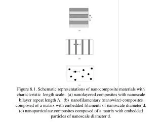
Figure 8.1. Schematic representations of nanocomposite materials with characteristic length scale: (a) nanolayered composites with nanoscalebilayer repeat length L; (b) nanofilamentary (nanowire) composites composed of a matrix with embedded filaments of nanoscale diameter d; (c) nanoparticulate composites composed of a matrix with embedded particles of nanoscale diameter d.
Figure 8.2. Schematic energy band diagram of GaAs/GaAlxAs1-x quantum well. An electron (represented by its wavefunction y) can be considered as partially confined in the quantum well of width equal to the GaAs thickness. The barrier height DE is equal to the difference in the energies of the bottom of the conduction band Ec for the two layer materials. Ev is the energy of the top of the valence band and Egap is the band gap energy.
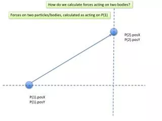
Figure 8.3. Precipitate particles of spacing l acting as obstacles to dislocation motion.
Figure 8.4. High resolution transmission electron micrograph showing a cross-sectional view of an InAs-GaSb (100) superlattice (Reproduced with kind permission of M. Twigg.)
Figure 8.5. Scanning electron micrograph of electrodeposited FeCo nanowires (the polycarbonate matrix in which the wires were embedded has been completely dissolved).
Figure 8.6. Bright field transmission electron micrographs of Ni/SiO2 granular metal films. (From Ref. 29 by permission of Elsevier Science B.V.)
Figure 8.7. Transmission electron micrographs of binary nanoparticle assemblies. (a) Fe3O4(4nm)-Fe58Pt42 (4nm) assembly; (b) Fe3O4 (8nm)- Fe58Pt42 (4nm) assembly; Fe3O4 (12 nm)- Fe58Pt42 (4 nm) assembly. (From Ref. 46 by permission of Macmillan MagazinesLtd.)