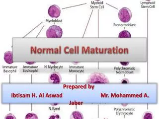
Normal Cell Maturation
Practical Hematology Lab. - LAB 12 -. Normal Cell Maturation. Blood Cells Maturation. Blood cells go through several stages of development; progression from one stage to the next is not rapid, so frequently the cell being studied may be between stages.

Normal Cell Maturation
E N D
Presentation Transcript
Practical Hematology Lab - LAB 12 - Normal Cell Maturation
Blood Cells Maturation • Blood cells go through several stages of development; progression from one stage to the next is not rapid, so frequently the cell being studied may be between stages. • When this occurs the cell is generally given the name of the mature stage). • As a cell transforms from the primitive blast stage to the mature form found in the blood, there are changes in the cytoplasm, nucleus, and cell size.
Normally, all three of these changes occurs gradually and simultaneously. • In some disease states, however, changes will take place at different rates
Cytoplasmic Maturation • The immature cytoplasm usually stain a deep blue color (basophilic) because of the high content of RNA present as the cell matures, there is a gradual loss of cytoplasmic RNA and therefore a lessening of blue color. • In some cells (for example, the myelogenous cells), granules appear in the cytoplasm as the cell matures. At first, these granules are few and nonspecific. As the cell matures further, these granules increase in number and take on specific characteristics and functions. • The amount of cytoplasm in relationship to the rest of the cell usually increases as the cell matures.
Nuclear Maturation • The nucleus of the immature cell is round oval and is large in proportion to the rest of the cell. As the cell matures the nucleus decreases in relative size and may or may not take on various shapes (depending on the cell type). • The nuclear chromatin transforms from a fine, delicate pattern to become more coarse and clumped on the mature form, and the staining properties change from a reddish purple to a bluish purple. • Nucleoli present in early stages of cell development usually disappear gradually as the cell ages.
Cell Size • As a cell matures, it usually becomes smaller in size. (for the new student, this change may be difficult to detect. • The normal mature red blood cells or small lymphocytes are generally of relatively constant size and may be used as a guide for comparison) the student should know the relative size of each cell type.
Identification of cells • In identifying a cell we should think of the following terms: • What is the size of the cell? • Small • Medium • Large • What are the characteristics of the cytoplasm? • Granular or nongranular; specific or nonspecific granules. • Color (staining properties). • Relative amount.
What are the characteristics of the nucleus? • Shape. • Relative size. • The size of the nucleus compared to the amount of cytoplasm is expressed as the nuclear to cytoplasmic ratio (N/C ratio). • Chromatin pattern: smooth or coarse. • Presence of or absence of nucleoli; number and size.
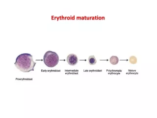
Erythrocyte Maturation The overall trend in RBC maturation is large, pale nucleus to darker, smaller nucleus to loss of nucleus; increase in cytoplasm; gradual decrease in size; cytoplasm from intensely blue (full of RNA) to grayish (mixture of RNA and hemoglobin) to reddish (full of hemoglobin, no RNA). Identify the following cells. Maturation
Erythrocyte Maturation • 1. Pronormobast (Rubriblast) • 14-19 µm • Nucleus is large with fine chromatin and nucleoli • Cytoplasm is scant and basophilic.
2. Basophilic Normoblas (Prorubriblast) • 12-17 µm • Slightly smaller nucleus with slight chromatin condensation • Increased cytoplasm and intensely blue (RNA abundance); no granules and no nucleoli present.
3. Polychromatophilic Normoblast (Rubricyte) • 12-15 µm • Moderately condensed chromatin • Lighter, grayish cytoplasm. • The color of the cytoplasm is due to coloring by both acidic and basic components of the stain. • Basophilia is from staining of ribosomes and acidophilia from hemoglobin. • The nucleus is condensed and intensely basophilic with coarse heterochromatin granules .
4. Orthochromatophilic Normoblast (Metarubricyte) • 8-12 µm • Dark, opaque nucleus; gray-red cytoplasm (trace blue). • The nucleus has become pyknotic (a homogenous blue-black mass with no structure) and there is abundant acidophilic hemoglobin. • In some instances you can detect the nucleus in the process of extrusion.
5.Reticulocyte: (7-10 µm) [Not visible with this preparation. Refer to the laser disc to see an example. Nucleus has been extruded; cytoplasm is reddish-pale blue. RNA is still present. 6.Erythrocyte: (7-8 µm): No nucleus; orange-red cytoplasm; RNA is lost.
Myeloid Maturation myeloblast promyelocyte myelocyte metamyelocyte band neutrophil MATURATION
1. Myeloblast • The first and earliest granulocyte • Is a large cell (15 μm) • High nucleus to cytoplasm (N:C) ratio (5:1) • Round or oval nucleus with loose light staining euchromatin • 1-2 nucleoli • Has minimal light blue cytoplasm • Contains no cytoplasmic granules • Begins to produce myeloperoxidase granules (MPO) • Comprises 1% of the nucleated cells in the bone marrow
2. Promyelocyte • Larger than a myeloblast (20 μm) • High N:C ratio (3:1) • Loose chromatin with nucleoli • Dark blue cytoplasm • Contains large nonspecific cytoplasmic granules • Containing myeloperoxidase (MPO) • Comprises 3-4% of nucleated bone marrow cells
3. Neutrophilic Myelocyte • Medium cell size (12 μm) • High N:C ratio (3:1) • Round, oval, or slightly indented nucleus with darker • Blue heterochromatin • Last stage of cell division • Has active RNA, therefore, the cytoplasm is blue • Contains MPO and secondary granules containing leukocyte alkaline phosphatase • Comprises 12% of bone marrow nucleated cells
4. Neutrophilic Metamyelocyte • 11 μm • N:C ratio (2:1) • Last mononuclear stage, no mitosis • Nucleus is kidney or horseshoe shaped, and has condensed heterochromatin • Has a prominent Golgi apparatus – clear area located at the indentation site of the nucleus • Cytoplasmis similar to the mature cell • Comprises 18% of bone marrow cells
5. Band Neutrophile • Same size as a mature neutrophil (10-12 μm) • N:C ratio has reversed (1:2) • Nucleus is band- or sausage-shaped without segmentation • Cytoplasm is filled with small neutrophilic granules • Last immature stage • Comprises 11% of bone marrow cells and 0-3% of peripheral WBCs Stored in the bone marrow and released when there is an increased demand for neutrophils • Shift to the left is an increase in immature cells indicating increased demand for WBCs in peripheral blood
1. Neutrophils • Also known as segmented neutrophils, segs, polymorphonuclear cells, polys, and PMNs • N:C ratio is 1:3, and the size is 10-12 μm • Average nucleus contains 3-5 segments connected by narrow filaments • Hyposegmented is less than 3 segments, and may indicate a shift to the left or an anomaly • Hypersegmented is more than 5 segments and may indicate infection or megaloblastic anemia • Cytoplasm contains very small neutral granules • Makes up 55-75% of all peripheral WBCs • Granules can become larger upon bacterial infection producing toxic granulation, which are numerous, large, basophilic granules
2. Eosinophils • Average size is 13 μm • Nucleus is generally bilobed • Cytoplasm is bright red or orange which is due to large specific, secretory granules containing peroxidase, acid phosphatase, aryl sulfatase, beta-glucuronidase, etc. that stain red with the eosin component of Wright’s stain • Makes up 3% of WBCs in the peripheral blood
3. Basophils • Is the smallest granulocyte at 10 μm • The nucleus is difficult to see due to heavy granulation • Cytoplasm contains large specific, secondary granules that contain heparin and histamines, which stain purple with Wright’s stain. • These granules are water soluble and sometimes appear as holes in the cell if the cells are not fixed well during staining. • Makes up to 0.5% of peripheral WBCs • Note: Tissue mast cells are similar to basophils but are larger and have no developmental relationship with basophils. Mast cells have a mesenchymal (connective tissue) origin and have granules containing serotonin (basophilic granules contain no serotonin).
4. Lymphopoiesis • A. Lymphoblast • Size : 10 to 18 µm • Cytoplasm : no granules present. Appears smooth. Moderate to dark blue. May stain deep blue at the periphery and a lighter blue near the nucleus. More abundant than in the myeloblast. • Nucleus : chromatin pattern is somewhat coarse. Round or oval in shape. • Generally contain one to two distinct nucleoli. • N/C ratio is 4:1.
B. Prolymphocyte • Size: May be the same size as the lymphoblast or smaller. • Cytoplasm: Moderate to dark blue. • Usually nongranular, but may contain occasional azurophilic granules. More abundant than in the lymphoblast. • Nucleus: Round, oval, or slightly indented. Chromatin pattern is more clumped than in the lymphoblast. May contain one to two nucleoli.
C. Small Lymphocyte • Size : 7 to 18 μm • N:C : 4:1 • Chromatin : Oval nucleus with coarse lumpy chromatin with specific areas of clumping, a compact cell • Cytoplasm : Usually just a thin border, with few azurophilic, red granules.
D. Large Lymphocyte • Size :9 to 12 μm • N:C : 3:1 • Chromatin : Looser chromatin pattern, more transparent • Cytoplasm : Larger amount of cytoplasm, lighter in color • Distinguishing characteristic :Cytoplasm is more abundant with tendency for azurophilic granules.
5. Monopoiesis • A. Monoblast • Size:12 to 20 µm • Cytoplasm: moderately basophilic to blue-gray. Nongranular. • Nucleus: Ovoid or round in shape. Light blue-purple in color. Fine, lacey chromatin. One to two nucleoli. • N/C ratio is 4:1 to 3:1.
B. Promonocyte • (immature monocyte) • Size: 14 to 18 µm • Cytoplasm : Blue-gray. May contain fine dustlike azurophilic granules. Ground glass appearance. Moderate amount. • Nucleus: oval, may have a single fold or fissure. One to five nucleoli. Fine chromatin pattern. • N/C ratio is 3:1 to 2:1.
C. Monocyte • Size: 14 to 20 µm • Cytoplasm:Abundant. Blue-gray. Outline may be irregular because of the presence of pseudopods. Many fine azurophilic granules, giving a ground glass appearance. Vacuoles may some times be present. • Nucleus: Round, kidney shaped, or may show slight lobulation. It may be folded over on top of itself, thus showing brain-like convolution. No nucleoli are visible. Chromatin is fine and lacey (not clumped), arranged in skin-like strands.
6. Megakaryocytopoiesis • A. Megakaryoblast • (stage 1) • Cytoplasm:varying shades of blue usually darker than the myeloblast. • May have small, blunt pseudopods. • Small to moderate amount. usually a narrow band around the nucleus. As the cell matures, the amount of cytoplasm increases. Usually nongranular.
Megakaryoblast (stage 1) • Nucleus: Round, oval, or may be kidney shaped. • Fine chromatin pattern. • Multiple nucleoli that generally stain blue. • N/C ratio is about 10:1.
B. Promegakaryocyte (stage 2) • Cytoplasm: more abundant than previous stage. • Lass basophilic than in the blast. Granules begin to form in the Golgi region. • Nucleus : chromatin becomes more coarse. Multiple nucleoli are visible. Irregular in shape; may even show slight lobulation. • N/C ratio is 4:1 to 7:1 depending on the ploidy.
C. Granular Megakaryocyte (stage3) • Cytoplasm : abundent. Pinkish blue in color. Very fine and diffusely granulat. Usually has an irregular peripheral border. • Nucleus : small in comparison to cell size. Multiple nuclei may be visible or the nucleus may show multilobulation. • Chromatin is coarser than in the previous stage. No nucleoli are visable. N/C ratio is 2:1 to 1:1.
D. Mature Megakaryocyte • (stage IV) • Cytoplasm: contain coarse clumps of granules aggregating into little bundles, which bud off from the periphery to become platelets. • Nucleus: multiple nuclei are present, or the nucleus is multilobulated. No nucleoli visible. N/C ratio is less than 1:1.
E. Platelet (Thrombocyte) • Size: 1 to 4 µm in diameter. • Cytoplasm: light blue to purple. Very granular. Consists of two parts: • The chromomere, which is granular and located centrally, and • The hyalomere, which surrounds the chromomere and is nongranular and clear to light blue.
Platelet satellitism: Platelets adhering to the surface of a neutrophil. In this case, the reported platelet count was only slightly decreased.