Introduction to Cell Biology: Microscope Basics and Cell Structures
560 likes | 622 Vues
Learn how to use a microscope and explore cell structures with this engaging starter activity. Discover the functions of cell components by examining onion and cheek cells. Dive into the world of cells, including plant and animal cell comparisons.

Introduction to Cell Biology: Microscope Basics and Cell Structures
E N D
Presentation Transcript
UNIT ONE CELL BIOLOGY NATIONAL 4 BIOLOGY
Starter Activity: Answer the following question in your jotter, in sentences Name three types of cells in your body. Approximately how many cells are there in the human body? How many cells are there in an Amoeba?
Answers • Blood, nerve, muscle, bone, brain, liver, skin etc • 100 trillion (100 million million) • One
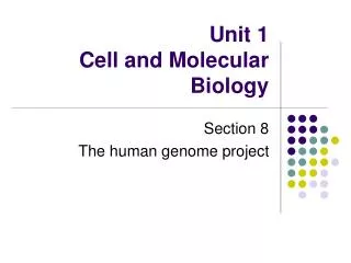
Key area 1 Cell structure and cell division
Learning Intentions • To know that a microscope can be used to magnify cells • Identify the parts of a microscope • Be able to set up and use a microscope to view different cells
Cells Cells are very small and they cannot be seen by the naked eye. We need to use a microscope to help us see them. A microscope contains special lenses to magnify cells.
Parts of a Microscope Eyepiece Lens Coarse Focus Fine Focus Arm Objective Lens Stage Clip Stage Base Mirror
How to use a microscope Carry the microscope using two hands. Plug it in and switch it on. Put the lowest power lens in place (the smallest one). Using the roughfocus wheel, move the stage away from the lens. Place the slide on the stage and hold it with the slide clips. Look at the microscope from the side. Turn the rough focus wheel until the slide is very close to the lens, but not touching it. (It should stop automatically)
Look down the microscope. Turn the rough focus so the slide moves downwards. • Stop when the slide is in focus. You may need to adjust it using the fine focus. • Now move the medium power lens into place. Use the fine focus wheel to re-focus it. • When you have finished, return the slide and pack away the microscope carefully.
Using a microscope to look at everyday objects • Look at each of the slides in turn. • In your booklet make drawings of what you see in the circles. • Give each drawing a title, and say what magnification you used (either x 40, x 100 or x 400). • E.g cotton wool x 100
Animal and plant cells Learning Intention: To review cell structure and function Success Criteria: Be able to name the structures common to animal and plant cells Be able to name the structures found in plant cells only Be able to label these on a diagram To be able to state the functions of these structures
Examining Onion Cells Equipment: • Glass slide • Cover slip • Onion skin • Iodine stain • Microscope • Lamp Aim: To observe and draw onion cells using a microscope.
Method: Collect a thin piece of onion skin. Spread the skin on a slide. The skin must not overlap. Stain the cells by adding 2 drops of iodine stain. Place a cover slip over the skin. Use a pencil to lower the cover slip gently so the air is pushed out. Examine the cells under low then medium power. You should be able to see lots of cells arranged like bricks in a wall. Adjust the microscope to a higher power. Draw exactly what you see through the “field of view” using a pencil. Label as many structures as you can see. Return the slide and pack your microscope away carefully.
Onion cells in iodine nucleus cell wall cytoplasm
A Typical Plant Cell cell membrane vacuole nucleus cell wall green chloroplast cytoplasm
Examining Cheek Cells Equipment: • Glass slide • Cover slip • Cotton bud • Methylene blue stain • Microscope and lamp • Paper towel Aim: To make a slide of cheek cells and draw them.
Method: Rub the cotton bud over the inside of your cheek to remove some of the cells. Wipe the cotton bud over the surface of a glass slide. Place the cotton bud in disinfectant. Stain the cells with 2 drops of methylene blue stain. Remove some of the stain using paper towel. Use a mounted needle to lower the cover slip so the air is pushed out. Draw the cells and label the structures. Once you have finished, place the slide and cover slip in disinfectant. Pack away your microscope carefully.
Cheek cells in methylene blue nucleus cell membrane cytoplasm
Typical Animal Cell nucleus cytoplasm cell membrane
Functions of cell structures • Each of these cell structures has a specific job to do in the cell. • This is called its function. • Do you know the functions of any of the cell structures?
Nucleus • The nucleus contains the genetic material of an organism. • It controls the cell’s chemical reactions. • It also controls the growth and development of a cell. nucleus
Cell membrane • Cells take in many chemicals from their surroundings, and release other chemicals into their surroundings. • The cell membrane is a very thin boundary which controls the entry and exit of these materials.
Cytoplasm • There are many chemical reactions happening in all of your cells. They happen in the cytoplasm. • These reactions keep the cell alive and allow it to carry out its specific function.
Cell wall • The cell wall is a rigid structure made of a tough mesh of cellulose fibres. • It helps to support a plant cell.
Vacuole • The vacuole is filled with water and pushes out towards the cell wall. • This provides support for the plant.
Chloroplasts • Plant cells may also contain chloroplasts in the cytoplasm. • These contain a chemical called chlorophyll which absorbs light energy for photosynthesis. • This allows plant cells to make food. • Only the green parts of a plant contain chloroplasts.
Try the matching cards2. Complete the table in your booklet Answers on next slide
Cell division Learning Intention: To understand the process of cell division Success Criteria: Be able to state the importance of cell division to living organisms Be able to describe the process of cell division
Importance of cell division pollen grain • Every living organism produced by sexual reproduction starts life as one cell. • In plants, this cell is formed when an egg cell is fertilised by a pollen grain. • The fertilised egg cell then divides into many cells to form a seed. • The seed can then grow into a new plant. pollen tube pollen nucleus egg cell
Importance of cell division • In animals, this cell is formed when an egg cell is fertilised by a sperm cell. • The fertilised egg then divides into many cells to form an embryo. • The embryo continues to grow into a new animal.
Growth • All living organisms grow during their lifetime. • All of this growth happens due to cells dividing to form new cells.
Repair to tissues • When we get injured, new cells are made to repair and replace the damaged tissues. • New skin cells form to heal a cut or graze, and new bone cells grow to repair a broken bone.
Cell division Learning Intention: Investigate the process of cell division Success Criteria: Be able to describe what happens during cell division Be able to describe the cells produced from cell division Be able to explain how cancer occurs
Starter Activity: On a Show Me Board, answer the following questions: Name the three main parts found in both animal and plant cells. Name two other structures which are found in plant cells only. Why are your cells dividing just now?
Cell division • Cells are able to make new cells by cell division. • The parent cell splits to form two cells. • Each new cell is identical to the original parent cell. Embryonic cell division
Cells and chromosomes • Inside the nucleus of a cell there are chromosomes which carry the genetic information of the organism. • Human cells have 46 chromosomes in their nucleus.
Chromosomes and cell division Chromosomes duplicate • Each new cell ends up with the same number of chromosomes as the parent cell. • This means each new cell has all the genetic information of the original cell. Two identical cells Chromatids separate
Revision checktest • One part found in all cells is the cytoplasm. Name the two other parts found in all cells. • Plant cells have a thick structure around the outside for support. What is it called? • What are the green structures in plant cells called? • What name is given to the structures in the nucleus of a cell which carry genetic information? • How many of these are there in human cells? • Your cells are able to divide to make new cells. Give a reason why cells in your body need to divide.
Cell division in bacteria • Bacteria are single celled living organisms. • They are too small to see with the naked eye. • Bacterial cells can divide very rapidly to form new cells. • When they do this, they form colonies of millions of cells which can be seen with the naked eye. • Your teacher will show you how to grow bacteria on an agar plate like the one shown in the bottom picture.
Growing bacteria on an agar plate Equipment : Sterile petri dish with agar Broth culture of bacteria Inoculating loop Bunsen burner Disinfectant Sticky tape Label Method : • Wash your hands and sterilise the bench with disinfectant. • Label the petri dish at the edge with your initials. • Heat the inoculating loop in a blue flame until it glows red hot. Allow it to cool for 20 seconds. • Dip the loop into the broth and remove. Replace the lid on the broth quickly. • Take the lid off the petri dish and gently spread the liquid onto the surface of the agar. • Replace the lid on the petri dish as quickly as possible. • Put the loop back into the flame until it glows red hot again. • Seal the dish with two pieces of sticky tape at opposite sides. • Sterilise the bench with disinfectant and wash your hands.
Growing bacteria on an agar plate • Results : Make a drawing of your petri dish after it has been incubated in an oven for 2-3 days. (Do not open the dish!)
Bacteria all around us • We are surrounded by bacteria all the time. • They can be found on your skin, on surfaces like your desk, in water, in the air – in fact they are everywhere! • Most of the bacteria around us are harmless, but some can be harmful. We sometimes call these harmful bacteria germs.
Bacteria all around us • Your teacher will show you how to use a swab (cotton bud) to collect bacteria from different places in the classroom. • You will grow these bacteria on agar in petri dishes. • After 3 days you will look at the results. • It is very important to keep the dishes fully sealed when looking at them as there may be harmful bacteria growing in the dish.
Bacteria all around us • It is important to protect ourselves from becoming ill due to these harmful bacteria. • Write down three ways in which you can avoid infection by harmful bacteria at home.
Avoiding infection at home • Wash hands before handling food. • Wash hands after using the toilet. • Keep kitchen surfaces and utensils clean. • Keep fresh food in a fridge. • Check sell-by dates on food. • Cook meat properly, especially chicken and pork. • Store cooked meat away from raw meat. • Cover left over food and store in the fridge. • Did you have any others?
Cell division Learning Intention: Investigate the process of cell division Success Criteria: Be able to explain how cancer occurs Be able to describe how limbs and organs can regenerate