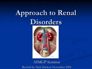
RENAL DISORDERS
RENAL DISORDERS. NUR -224. Objectives. Differentiate between the causes of acute and chronic kidney failure. Describe the nursing management of patients with acute and chronic renal failure. Compare and contrast renal replacement therapies. Glomerulus.

RENAL DISORDERS
E N D
Presentation Transcript
RENAL DISORDERS NUR -224
Objectives • Differentiate between the causes of acute and chronic kidney failure. • Describe the nursing management of patients with acute and chronic renal failure. • Compare and contrast renal replacement therapies.
Glomerulus • Glomerulus is a tuft of capillaries surrounded by the Bowman’s capsule. • The amount of fluid filtered from the blood into the capsule /min is called the glomerular filtration rate • 99% of the filtrate is reabsorbed in the renal tubules. • Glomerular disease affects both the structure and function of the glomerulus & is the leading cause of CKD in the US.
Glomerular Disease • Glomerular disease affects both the structure and function of the glomerulus disrupting glomerular filtration. • Capillary membrane becomes more permeable to plasma proteins and blood. • This increased permeability causes hematuria, proteinuria, and edema. • As the GFR falls, azotemia
Glomerular Disease • Loss of plasma proteins leads to hypoalbuminemia edema. • As plasma proteins are lost, filtration increases. • This stimulates renin-angiotensin-aldosterone mechanism vasoconstriction & salt/water retention • As the GFR continues to fall oliguria and hypertension
ACUTE GOLMERULONEPHRITIS • Inflammation of the glomeruli capillary membrane • Results from systemic diseases/primary glomerular diseases • Post-streptococcal glomerulonephritis – most common • Antigen/antibody complexes form during the primary infection and become trapped in glomerular capillaries inflammatory response
ACUTE NEPHRITIC SYNDROME Risk factors • Impetigo • Group A beta-hemolytic streptococcal infection • Acute viral infections • Hepatitis B • Primarily a childhood disease
ACUTE NEPHRITIC SYNDROME Clinical manifestations • Hematuria • Edema –low pressure areas • Azotemia • Proteinuria • Hypertension Prognosis less favorable for adults.
ACUTE NEPHRITIC SYNDROME Assessment/Diagnostic Findings • Throat/skin cultures • KUB abdominal x-ray • Serum BUN /creatinine levels • Urinalysis • Kidney biopsy
ACUTE NEPHRITIC SYNDROME Medical Management – Medications • no medication available to cure the disease • used to treat the underlying cause, reduce inflammation, manage the symptoms • Antibiotics • Oral glucocorticoids • Antihypertensive
ACUTE NEPHRITIC SYNDROME Nursing Management / Excessive Fluid volume • Monitor vital signs • Record intake/output • Maintain fluid restriction as ordered • Monitor for desired effects of prescribed medication • Provide frequent position changes • Arrange dietary consultation
CHRONIC GLOMERULONEPHRITIS • Repeated episodes of acute nephritic syndrome • Kidneys reduced to one-fifth of their normal size. • Glomeruli/tubules become severely scarred • Reflects the end-stage of glomerular inflammatory disease. • May require renal transplant
CHRONIC GLOMERULONEPHRITIS • May have no symptoms for many years General symptoms – • loss of weight, increasing irritability, nocturia, • gray pigmentation • periorbital/peripheral edema
CHRONIC GLOMERULONEPHRITIS Assessment/Diagnostic Findings • electrolyte changes • anemia • CXR, CT-scan • Mental status changes
CHRONIC GLOMERULONEPHRITIS Medical management • Symptoms guide the treatment. • Manage hypertension • Diet/weight • Dialysis
KIDNEY FAILURE • Results when the kidneys cannot remove wastes or perform regulatory functions • A systemic disorder that results from many different causes • Acute kidney failure is a reversible syndrome that results in decreased GFR and oliguria • Chronic kidney failure (ESRD) is a progressive, irreversible deterioration of renal function that results in azotemia
ACUTE KIDNEY FAILURE • Rapid loss of renal function due to damage of the kidneys • Slight deterioration in kidney function severe impairment • Increase in serum creatinine • Decrease in urine output • Development of azotemia
ACUTE KIDNEY FAILURE (AKI) Pathophysiology • May be a specific underlying problem problem that will reduce blood flow to the kidneys • Conditions should be treated and corrected before there is permanent damage to the kidneys.
Categories of Acute Renal Failure • Prerenal • Intrarenal • Postrenal
PRERENAL • factors external to the kidneys • occurs in 60-70% of cases • factors reduce systemic circulation reduction in renal blood flow decrease glomerular perfusion • if condition persists kidneys lose their ability to compensate causing damage to renal tissues. • dehydration, excessive diuresis, heart failure, neurologic injury, burns
INTRARENAL • Cause direct damage to the kidney tissue impair nephron function • ATN/acute tubular necrosis – common cause • Caused by prolonged ischemia or nephrotoxins – responsible for 90% of intrarenal cases • Nephrotoxins – obstruction of intrarenal structures – slough off and plug the tubules
POSTRENAL • Involve mechanical obstruction in urine outflow • As urine flow is obstructed - urine refluxes into the renal pelvis impaired renal function • Causes – BPH, prostate cancer, calculi, tumors • Post renal causes of AKI account for less than 10% of AKI cases.
CLINICAL MANIFESTATIONS • Prerenal/postrenal usually resolve quickly with correction of the cause. • AKI has a prolonged course – progress through phases. • Phases: initiation oliguric diuretic recovery
OLIGURIC PHASE • Increase in serum concentration of substances usually secreted by the kidneys • Urine output needs to be at least 400mL/day • Oliguria – most common manifestations • Fluid volume • Hyperkalemia • BUN/Creatinine • Neurologic status
DIURETIC PHASE • Gradual increase in urine output – 1-3L/day, nephrons may still not be fully functional • Lasts from 1-3 weeks • Kidneys have recovered their ability to excrete waste but not to concentrate urine • Monitor for hypervolemia, hypotension
RECOVERY PHASE • Improvement of renal function (may take several months) • Lab values return to normal
ASSESSMENT/DIAGNOSTIC FINDINGS Prerenal – dehydration, blood loss, heart disease Intrarenal – nephrotoxic drugs, hx of blood transfusions, radio opaque contrast media Postrenal- stones, BPH, cancer - bladder/prostate • Ultrasonography • Urinalysis • Renal Biopsy
Acute Renal Failure Medical management • Eliminate the underlying cause • Maintain fluid balance • Avoid fluid excesses – diuretic therapy • Renal replacement therapy (RRT) prevent complications hemodialysis – peritoneal dialysis continuous renal replacement therapy
NUTRITIONAL THERAPY • Goal – maintain adequate calorie intake • Should come from CHO and fat sources • Sodium should be restricted to prevent edema • May need enteral nutrition feedings • Fat emulsions IV may be given as a source of nonprotein calories
Acute Renal Failure Nursing management • Fluid/electrolyte balance • Prevent infection • Provide skin care • Provide psychosocial support
Chronic Renal FailureESRD/CRD • Involves progressive, irresistible loss of kidney function. • RRT/transplantation is required at this point • Presence of kidney damage for more than 3 or more months • Leading cause – diabetes (2/3 of cases) and hypertension making up 1/3 of the cases • Incidence of CRD is significantly higher in people over 65.
Chronic Renal Failure Pathophysiology • Renal function declines end products of protein metabolism accumulate in the blood. • Uremia develops affects every system in the body
CHRONIC RENAL FAILUREESRD Clinical Manifestations • s/s depend on degree of renal impairment and every body system is affected • Chart – pg. 1326
Assessing ESRD • Neuro – • Skin – • CV - • Pul - • GI – • Musculo –
Chronic Renal Disease Assessment/ Diagnostic Findings • Metabolic disturbances • Electrolyte /acid-base imbalances • Anemia • Calcium/phosphorus imbalance
Lab Data Metabolic disturbances • GFR decreases creatinine/BUN levels increase • Uremia • Diabetics may require less insulin than before the onset of CRF
Lab Data Electrolyte/acid-base imbalances • Hyperkalemia –serious electrolyte disturbance • Sodium – normal or low in kidney failure • Causes edema, heart failure, hypertension • Metabolic Acidosis -kidneys are unable to excrete acid • Kidneys are unable to excrete ammonia and reabsorb sodium bicarbonate
Lab Data Anemia • Inadequate erythropoietin production • Erythropoietin normally produced by the kidneys, stimulates bone marrow to produce RBC • ESRD decrease production of erythropoietin leads to anemia fatigue, angina, SOB
Lab Data CKD mineral/bone disorder • Kidney function deteriorates, less Vitamin D is converted to its active form decreased serum levels • Vitamin D is necessary for calcium to absorbed from the GI tract • Decrease Vitamin D levels decrease in calcium from the intestines decrease serum calcium levels
Lab Data • CKD mineral/bone disorder • Hypocalcemia occurs parathyroid gland secretes parathyroid hormone (PTH) stimulates bone demineralization releasing calcium from the bone. • Phosphate is released as well - hyperphosphatemia –decrease phosphate excretion by the kidneys • Low calcium, high phosphate levels and decreased Vitamin D – parathyroid – PTH – bone remodeling – risk for fractures
MEDICAL MANGEMENT Goal – • Maintain kidney function and homeostasis for as long as possible • All factors that are reversible are identified and treated • Management accomplished – medication and diet therapy
PHARMACOLOGIC THERAPY Complications are prevented or delayed by administering : • Calcium/Phosphate-binding agents • Antihypertensive and cardiac medications • Anti-seizure medications • Erythropoietin
Nutritional Therapy • Protein restriction • Fluid restriction • Sodium restriction • Vitamin supplement • Monitor foods high in phosphate
Renal Replacement Therapy • Becomes necessary when the kidneys can no longer remove wastes, maintain electrolytes and regulate fluid balance • Various types of dialysis and kidney transplant
Dialysis • Hemodialysis – circulates the patient’s blood through a dialyzer to remove waste products and excess fluid • Peritoneal dialysis – uses the patient peritoneal membrane as the semi-permeable membrane to exchange fluid and solutes • Continuous renal replacement therapy – method used to replace normal kidney by circulating the patient’s blood through a hemofilter