Obstructive Renal Disorders
Obstructive Renal Disorders. NPN 200 Medical Surgical Nursing I. Hydronephrosis, Hydroureter, and Urethral Stricture. Outflow obstruction Urethral stricture Causes bladder distention and progresses to the ureters and the kidneys Hydronephrosis –


Obstructive Renal Disorders
E N D
Presentation Transcript
Obstructive Renal Disorders NPN 200 Medical Surgical Nursing I
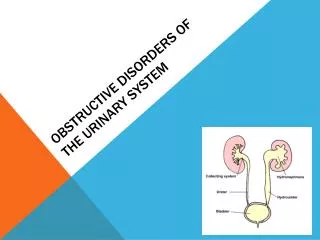
Hydronephrosis, Hydroureter, and Urethral Stricture • Outflow obstruction • Urethral stricture • Causes bladder distention and progresses to the ureters and the kidneys • Hydronephrosis – • Kidney enlarges as urine collects in the pelvis and kidney tissue due to obstruction in the outflow tract • Over a few hours this enlargement can damage the blood vessels and the tubules • Hydroureter • Effects are similar, but occurs lower in the ureter
Causes of Obstruction • Tumor • Stones • Congenital structural defects • Fibrosis • Treatment with radiation in pelvis
Complication of Obstruction • If untreated, permanent damage can occur within 48 hours • Renal failure • Retention of • Nitrogenous wastes (urea, creatinine, uric acid) • Electrolytes (K, Na, Cl, and Phosphorus) • Acid base balance impaired
Renal Calculi • Called nephrolithiasis or urolithiasis • Most commonly develop in the renal pelvis but can be anywhere in the urinary tract • Vary in size –from very large to tiny • Can be 1 stone or many stones • May stay in kidney or travel into the ureter • Can damage the urinary tract • May cause hydronephrosis • More common in white males 30-50 years of age
Renal Calculi • Predisposing factors • Dehydration • Prolonged immobilization • Infection • Obstruction • Anything which causes the urine to be alkaline • Metabolic factors • Excessive intake of calcium, calcium based antacids or Vit D • Hyperthyroidism • Elevated uric acid
Renal Calculi • Subjective symptoms • Sever pain in the flank area, suprapubic area, pelvis or external genitalia • If in ureter, may have spasms called “renal colic” • Urgency, frequency of urination • N/V • Chills
Renal Calculi • Objective symptoms • Increased temperature • Pallor • Hematuria • Abdominal distention • Pyuria • Anuria • May have UTI on urinalysis
Renal Calculi • Diagnostic procedures • Urinalysis with C and S • 24 hour urine • KUB • IVP • Renal CT • Kidney ultrasound • Cystoscopy with retrograde pyleogram
Renal Calculi • Treatment • Most are passed without intervention • May need cysto with basket retrieval • Lithotripsy • Lasertripsy • Lithotomy
Renal Calculi • Assessment • History and physical exam • Location, severity, and nature of pain • I/O • Vital signs, looking for fever • Palpation of flank area, and abdomen • ? N/V
Renal Calculi • Nursing interventions • Primary is to treat pain – usually with opioids • Ambulate • Force fluids, may have IV • Watch for fluid overload • Strain urine – send stone to lab if passed • Accurate I/O • Medicate N/V
Renal Calculi • Surgical removal • Routine pre and post op care • May return with catheter, drains, nephrostomy tube and ureteral stent – must maintain patency and may need to irrigate as ordered • Measure drainage from all tubes – need at least 30 cc/hr • Watch site for bleeding • May need frequent dressing changes due to fluid leakage, or may have collection bag
Renal Calculi • Discharge and prevention • Continue to force fluids post discharge • May need special diet • Stones are analyzed for calcium or other minerals • May need to watch products with calcium
Benign Prostatic Hypertrophy (BPH) • Enlargement of prostate gland which obstructs urinary out flow • Urinary stream decreases, with dysuria • Gradual dilation of ureters and kidney due to stasis • Age related and slow progressing, usually>50 • Cause is unknown – possible arteriosclerosis, changes in hormone levels, or inflammation
Subjective symptoms Urgency, frequency, burning, and hesitancy Decreased force of urination Nocturia Objective symptoms Voiding small amounts Hematuria Urinary retention Infection Enlarged prostate on exam Renal insufficiency Benign Prostatic Hypertrophy (BPH)
Benign Prostatic Hypertrophy (BPH) • Diagnostic tests • Client history • Physical exam • Residual cath • IVP • BUN and creatitine levels • UA and C&S • Cystoscopy
Benign Prostatic Hypertrophy (BPH) • Treatment • Drugs • Testosterone ablating agents – diethystilbestrol (DES) • Testosterone sparing – Proscar, which reduces the size of the gland • Alpha blockers – Flomax, relax smooth muscle in the bladder neck and prostate
Benign Prostatic Hypertrophy (BPH) • Surgical removal • TURP • Subrapubic, retropubic, or perineal prostectomy • Other methods to improve function • Sexual intercourse, hot sitz baths, or prostatic massage- which releases prostatic fluid pressure
Benign Prostatic Hypertrophy (BPH) • Post- op care routine + • With TURP will have a catheter, possibly 3-way for irrigation • Watch for blood clots • Keep accurate I/O • Give pain RX and antispasmotics • Encourage fluids
Benign Prostatic Hypertrophy (BPH) • Complications • At risk for UTI and retention • Erectile dysfunction • Hemorrhage • Urinary leakage • Sterility