Download
1 / 14
140 likes | 158 Vues
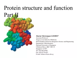
Protein structure and function Part II. Marie-Véronique CLEMENT Associate Professor Yong Loo Lin School of Medicine NUS Graduate School for Integrative Science and Engineering Department of Biochemistry National University of Singapore 8 Medical Drive, MD 7 #03-15 Singapore 117597

E N D
Protein structure and function Part II Marie-Véronique CLEMENT Associate Professor Yong Loo Lin School of Medicine NUS Graduate School for Integrative Science and Engineering Department of Biochemistry National University of Singapore 8 Medical Drive, MD 7 #03-15 Singapore 117597 Tel: (65) 68747985 Fax: (65) 67791453 E-mail: bchmvc@nus.edu.sg
TERTIARY STRUCTURE • R-group interactions result in 3D structures of globular proteins • Types of interactions : H-, ionic- (salt linkage), hydrophobic- and disulphide- bond • Hydrophilic R groups on surface while hydrophobic R groups buried inside of molecule • Wide variety of 3o structures: since large variation in protein sizes and amino acid sequences
The role of side chain in the shape of proteins Where is water? Hydrophilic Hydrophobic
Similarly: The tertiary structure for myoglobin is fairly well understood. Myoglobin has an alpha helix which then can be viewed as being enclosed in this blue sheath, the sheath doesn't exist but we can draw it that way. That helix folds back upon itself into what's referred to as the tertiary structure of myoglobin. Bonds between the side groups of the amino acid residues are responsible for holding together the tertiary structure of this protein.
A coiled-coil: Structure occurs when the 2 a helix have most of their nonpolar (hydrophobic) side chains on one side, so that they can twist around each other with these side chain facing inwards
Quaternery structure: If protein is formed as a complex of more than one protein chain, the complete structure is designed as quaternery structure: • Generally formed by non-covalent interactions between subunits • Either as homo- or hetero-multimers
QUATERNARY STRUCTURE: ADVANTAGES • Oligomers (multimers) are more stable than dissociated subunits • They prolong life of protein in vivo • Active sites can be formed by residues from adjacent subunits/chains • A subunit may not constitute a complete active site • Error of synthesis is greater for longer polypeptide chains • Subunit interactions : cooperativity/ allosteric effects
Primary structure Secondary structure Tertiary structure Quaternary structure