Download
1 / 28
280 likes | 536 Vues
BMS 631 - LECTURE 1 Flow Cytometry: Theory J.Paul Robinson Professor of Immunopharmacology Professor of Biomedical Engineering Schools of Veterinary Medicine & Engineering Purdue University. Introduction Course Requirements Lecture Series: 2002. Hansen Hall, B050 Purdue University

E N D
BMS 631 - LECTURE 1Flow Cytometry: TheoryJ.Paul RobinsonProfessor of ImmunopharmacologyProfessor of Biomedical EngineeringSchools of Veterinary Medicine & EngineeringPurdue University • Introduction • Course Requirements • Lecture Series: 2002 Hansen Hall, B050 Purdue University Office: 494 0757 Fax 494 0517 email\; robinson@flowcyt.cyto.purdue.edu WEB http://www.cyto.purdue.edu ©J.Paul Robinson, Purdue University BMS 631 - LECTURE1.PPT
Structure of this course • Lectures: The class consists primarily of lectures and lecture discussions with mini tutorials as necessary. • Practicals: There are no practical components to the 631 course. We will however, look at some instruments and instrument components to gain some perspectives. • Quizes: There is a midterm quiz and an end of term quiz. They are worth 60% of the grade. • Seminar: Each student must present a seminar at the conclusion of the course. This seminar must be discussed with the course director prior to preparation. This is worth 30% of the final grade. ©J.Paul Robinson, Purdue University BMS 631 - LECTURE1.PPT
Sources of information • Flow Cytometry and Sorting, 2nd ed. (M.R. Melamed, T. Lindmo, M.L. Mendelsohn, eds.), Wiley-Liss, New York, 1990 - referred to here as MLM • Flow Cytometry: Instrumentation and Data Analysis (M.A. Van Dilla, P.N. Dean, O.D. Laerum, M.R. Melamed, eds.), Academic Press, London, 1985 – referred to as VDLM • Practical Flow Cytometry 3nd edition (1994),H. Shapiro: Alan R. Liss, New York - referred to as PFC • Introduction to Flow Cytometry. J. Watson, Cambridge Press, 1991 referred to as IFC • Methods in Cell Biology: v.40,41, 63, 64 Darzynkiewicz, Robinson & Crissman, Academic Press, 1994, 2000 MCB • Data Analysis in Flow Cytometry:A Dynamic Approach-Book on CDROM M. Ormerod referred to as DAFC • Flow Cytometry: First Principles. (2nd Ed) Alice Longobardi Givan, Wiley-Liss, 2001 referred to as AFCFP More information on flow cytometry books can be found on our website at: http://www.cyto.purdue.edu/flowcyt/books/bookindx.htm Note: All of these books are in Prof. Robinson’s library in Hansen Hall, Room B50 and may be checked out for 24 hour periods with permission. ©J.Paul Robinson, Purdue University BMS 631 - LECTURE1.PPT
Sources • Flow Cytometry and Sorting, 2nd ed. (M.R. Melamed, T. Lindmo, M.L. Mendelsohn, eds.), Wiley-Liss, New York, 1990 - referred to here as MLM • Flow Cytometry: Instrumentation and Data Analysis (M.A. Van Dilla, P.N. Dean, O.D. Laerum, M.R. Melamed, eds.), Academic Press, London, 1985 - VDLM • Practical Flow Cytometry 4th edition (2003),H. Shapiro: Alan R. Liss, New York - PFC • Introduction to Flow Cytometry. J. Watson, Cambridge Press, 1991 IFC • Methods in Cell Biology: v.40,41, 62, 63 Darzynkiewicz, Robinson & Crissman, Academic Press, 1994, (Vol 62,63, 2000) MCB • Data Analysis in Flow Cytometry:A Dynamic Approach-Book on CDROM M. OrmerodDAFC • Flow Cytometry: First Principles. 2nd Edition Alice Longobardi Givan, Wiley-Liss, 2000 AFCFP ©J.Paul Robinson, Purdue University BMS 631 - LECTURE1.PPT
Methods and Practical Assistance • For help with protocols there are several choices including the MCB references on the previous slide (Methods in Cell Biology) • The Handbook of Flow Cytometry Methods • Current Protocols in Cytometry ©J.Paul Robinson, Purdue University BMS 631 - LECTURE1.PPT
Reference Material • The course will use Shapiro: • Practical Flow Cytometry, 4nd edition (2003), Howard Shapiro, Wiley-Liss, New York, as the main reference text. • Supplementary texts: • Introduction to Flow Cytometry. J. Watson, Cambridge Press, 1991 • Flow Cytometry: First Principles. Alice Longobardi Givan, Wiley-Liss, 1992 • Flow Cytometry: A Practical Approach. M.G. Omerod, IRL Press, 1990 • Methods in Cell Biology: vols 40,41. Darzynkiewicz, Robinson & Crissman, Academic Press, 1994 • Flow Cytometry, Advanced Research and Clinical Applications. A. Yen, CRC Press ©J.Paul Robinson, Purdue University BMS 631 - LECTURE1.PPT
Additional Sources • Powerpoint presentations references as J.Paul Robinson (JPR); Robert Murphry (RFM), Carleton Stewart (CS) • Web sources of these presentation are: • http://www.cyto.purdue.edu/flowcyt/educate/pptslide.htm • http://www.cyto.purdue.edu/flowcyt/educate1.htm Additional Sources include the Purdue Cytometry CD-ROM series Vol. 1 Vol. 2 Vol. 3 Vol. 4 Vol. 5 Microscopy 1 Volumes 6 and 7 (Cytomics) are also available as of 2002 and 2003 ©J.Paul Robinson, Purdue University BMS 631 - LECTURE1.PPT
Week 1 • Introduction to the course. • Discussion of texts and associated reading materials. • Discussion of expectations of students and special concerns. • Methods of evaluation and testing for the course. • Allocation of special review areas and discussion of areas for presentation of laboratory seminar. • Introduction to flow cytometry principles • References: (Shapiro pp 1-5; Watson pp 1-4; Givan pp 1-9) ©J.Paul Robinson, Purdue University BMS 631 - LECTURE1.PPT
Allowable Topics for Seminars The topic for the student seminar must be based upon an understanding of a component of the technology. It must demonstrate a complete understanding of the principle involved and the application to biology. Evaluation: The seminar counts for 30% of the course. See requirements below. EXAMPLES OF PREVIOUS SEMINARS - Evaluation of Small Particles using Flow Cytometry - Kinetic Measurements using Flow Cytometry - Monoclonal Antibodies, Avidin-Biotin Technology using Fluorescent Conjugates in Flow Cytometry - Fluorescent Molecules used in Flow Cytometry - Optical Filters used in Flow Cytometry - The Optical System in a Flow Cytometer - The Fluidic System of a Flow Cytometer - The Principles of Sorting in Flow Cytometry - Parameters used in Flow Cytometry - Parameters & Probes for Evaluation of DNA & RNA in Flow Cytometry - others will be added as necessary RULES: Presentations on research projects WILL NOT BE ALLOWED. The purpose of this seminar is to demonstrate your technical knowledge in a particular area of flow cytometry. The seminar may be videotaped and must not exceed 20 minutes or be less than 15 minutes. All presentations must be made using Powerpoint. Copies of both electronic and hardcopy must be provided in advance for evaluation. All material must be approved by the course instructor before presentation. Student Seminars ©J.Paul Robinson, Purdue University BMS 631 - LECTURE1.PPT
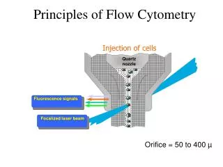
General introduction to flow cytometry • Introduction to the terminology, types of measurements, capabilities of flow cytometry, uses & applications • Comparison between flow cytometry and fluorescence microscopy • Transmitted light • Scatter • Sensitivity, precision of measurements, statistics, populations ©J.Paul Robinson, Purdue University BMS 631 - LECTURE1.PPT
Publications using the keyword “flow cytometry” from 62,496 references (2002) 1st The growth of diagnostic and phenotypic determination technologies ©J.Paul Robinson, Purdue University BMS 631 - LECTURE1.PPT
1400 • Papers Published per year in: • Image Cytometry • Image Analysis • Confocal Microscopy • Confocal } 1200 Medline 1000 800 Papers 600 400 200 0 1972 1970 1974 1976 1978 1980 1982 1984 1986 1988 1990 1992 1994 1996 1998 Year ©J.Paul Robinson, Purdue University BMS 631 - LECTURE1.PPT
Technical Components • Detection Systems Photomultiplier Tubes (PMTs) Historically 1-2 Current Instruments 3-9 Diodes Light scatter detectors • Illumination Systems Lasers (350-363, 405, 420, 457, 488, 514, 532, 600, 633 nm) Argon ion, Krypton ion, HeNe, HeCd, Yag Arc Lamps Mercury, Mercury-Xenon (most lines) ©J.Paul Robinson, Purdue University BMS 631 - LECTURE1.PPT
Stains... Up to 1850’s - only natural stains were available such as Saffron (which was what Leeuwenhoek used to stain muscle cells) Ehrlich - used acidic and basic dyes to identify acidophilic, eosinophilic, basophilic and neutrophilic leukocytes 1880’s to study the dynamics of ocular fluids- used fluorescein Fluorescence UV Microscope - August Köhler - 1904 Pappenheim & Unna (early 1900’s) - combined methyl green and pyronin to stain nuclei green and cytoplasm red Robert Feulgen (1925) - demonstrated that DNA was present in both animal and plant cell nuclei - developed a stoichiometric procedure for staining DNA involving a derivatizing dye, (fuchsin) to a Schiff base ©J.Paul Robinson, Purdue University BMS 631 - LECTURE1.PPT
Fluorescence Labeling Technique “Moreover, when Type II and III organisms were dried on different parts of the same slide, exposed to the conjugate for 30 minutes, washed in saline and distilled water, and mounted in glycerol, individual Type III organisms could be seen with the fluorescence microscope……” Coons et al 1941 - developed the fluorescence antibody technique - they labeled antipneumococcal antibodies with anthracine allowing them to detect both the organism and the antibody in tissue using UV excited blue fluorescence Manuscript: Immunological Properties of an Antibody Containing a Fluorescent Group Albert H. Coons, Hugh J. Creech and R. Norman Jones Department of Bacteriology and Immunology, Harvard Medical School, and the Chemical Laboratory, Harvard University Proc. Soc. Exp.Biol.Med. 47:200-202, 1941 Coons and Kaplan (1950) - conjugated fluorescein with isocyanate - better blue green fluorescent signal - further away from tissue autofluorescence. This method used a very dangerous preparative step using phosgene gas ©J.Paul Robinson, Purdue University BMS 631 - LECTURE1.PPT
Avery • (1944)Oswald T. Avery (1887-1955) - demonstrated that DNA was the carrier of genetic information The Discovery of the "Transforming Principle" Avery’s key discovery was that the transforming substance, which produced permanent, heritable change in an organism (pneumococci), was deoxyribonucleic acid. The phenomenon of transformation, Avery wrote, "has been interpreted from a genetic point of view. The inducing substance has been likened to a gene, and the capsular antigen which is produced in response to it has been regarded as a gene product." …. ”…If the results of the present study on the chemical nature of the transforming principle are confirmed, then nucleic acids must be regarded as possessing biological specificity...." Journal of Experimental Medicine, 1944 ©J.Paul Robinson, Purdue University BMS 631 - LECTURE1.PPT
Gucker - 1947 • Developed a flow cytometer for detection of bacteria in aerosols • Published paper in 1947 (work was done during WWII and was classified). • Goal was rapid identification of airborne bacteria and spores used in biological warfare • Instrument: Sheath of filtered air flowing through a dark-field flow illuminated chamber. Light source was a Ford headlamp, PMT detector (very early use of PMT) ©J.Paul Robinson, Purdue University BMS 631 - LECTURE1.PPT
Friedman Friedman (1950) - combined acid fuchsin, acridine yellow and berberine for uterine cancer detection using fluorescence microscopy Acid Fuchsin Other Names Acid magentaAcid rubinAcid roseine Absorption Max 540-545 ©J.Paul Robinson, Purdue University BMS 631 - LECTURE1.PPT
P.J. Crossland-Taylor “Provided there is no turbulence, the wide column of particles will then be accelerated to form a narrow column surrounded by fluid of the same refractive index which in turn is enclosed in a tube which will not interfere with observation of its axial content.” Sheath Flow Principle A Device for Counting Small Particles Suspended in a Fluid through a Tube P.J. Crosland-Taylor Bland-Sutton Institute of Pathology Middlesex Hospital, London, W.1. June 17, 1952 Nature 171: 37-38, 1953 ©J.Paul Robinson, Purdue University BMS 631 - LECTURE1.PPT
Watson & Crick J. D. WATSON and F. H. C. CRICKA Structure for Deoxyribose Nucleic Acid Nature, 2 April 1953, VOL 171,737 1953 The work which began with Avery's identification of DNA as the "transforming principle" thus led to research that overturned the old conception of DNA as a repetitive and simple molecule, confirmed DNA's role in genetic transmission, and, with James Watson and Francis Crick's 1953 paper, elucidated its structure. This is a picture of part of the original model built by Watson and Crick at Cambridge in 1953. ©J.Paul Robinson, Purdue University BMS 631 - LECTURE1.PPT
von Bertalanffy & Bickis • Ludwig von Bertalanffy (1901--1972) • von Bertalanffy & Bickis (1956) • - the metachromatic fluorescence of AO was used to identify and quantitate RNA in tissues • - also that normal and malignant cells could be discriminated Absorption Max 467 nm ©J.Paul Robinson, Purdue University BMS 631 - LECTURE1.PPT
©J.Paul Robinson Early Cell Counter Early cell counter. Katherine Williams and C.S. Sanders (Atomic Energy Research Establishment) 1948 - Unclassified in 1956. (Photo taken in Science Museum, London UK) ©J.Paul Robinson, Purdue University BMS 631 - LECTURE1.PPT
The first Coulter Counter High Speed Automatic Blood Cell Counter and Cell Size Analyzer Wallace H. Coulter Coulter Electronics, Chicago, Illinois :1034-1042, 1956 ©J.Paul Robinson, Purdue University BMS 631 - LECTURE1.PPT
©J.Paul Robinson Wallace Coulter - Coulter orifice - 1948-1956 Cell counter vacuum orifice
©J.Paul Robinson Hand-drawn advertising drafts of the first Coulter Counter ©J.Paul Robinson, Purdue University BMS 631 - LECTURE1.PPT
Historical Overview • IV. Requirements of image analysis • Pattern recognition, feature extraction, parameters or descriptors, texture Why was it so difficult to do image analysis and image processing in the 1960’s? ©J.Paul Robinson, Purdue University BMS 631 - LECTURE1.PPT
Historical Overview V. Early flow systems Hallermann et al, Kosenow - 1964 - AO staining of leukocytes - was able to use fluorescence (in a flow based system) to select leukocytes from red cells despite a low ratio (1/1000) because they took up lots of AO - also claimed to be able to discriminate between monocytes and PMN ©J.Paul Robinson, Purdue University BMS 631 - LECTURE1.PPT
Lecture Summary • Introduction to course • Reading and Support Materials • History • Technical Highlights • At the conclusion of this lecture you should: • Know what the requirements of this course are • Know where to track down information of importance • Understand a brief history of the development of flow technology • Be introduced into some of the principles • Have a perspective on why imaging was so difficult to do ©J.Paul Robinson, Purdue University BMS 631 - LECTURE1.PPT