Download
1 / 150
1.72k likes | 3.03k Vues
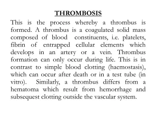
Thrombosis and embolism . By Dr S Homathy. Thrombosis. Thrombosis is the formation of a solid mass (blood clot) from the constituents of blood Platelets Fibrin Entrapped red cells and white cells Within the heart or vascular system in a living organism.

E N D
Thrombosis and embolism By Dr S Homathy
Thrombosis • Thrombosis is the formation of a solid mass (blood clot) from the constituents of blood • Platelets • Fibrin • Entrapped red cells and white cells • Within the heart or vascular system in a living organism
The development of a clot is life-saving when a large vessel ruptures or is severed. • However, when a thrombus develops within the vascular system, it may be life-threatening
Thrombosis is the consequence of inappropriate activation ( pathological) of the processes of normal haemostasis
Normal Haemostasis • Maintain blood in a fluid, clot-free state in normal vessels • Also inducing the rapid formation of a localized haemostatic plug at the site of injury. • Both are influenced by • components of the blood vessel wall, • platelets • the clotting sequence.
The integrity of the blood vessel wall is crucial in normal haemostasisas well as in thrombosis.
Normal Haemostasis Vessel injury – • brief period of arteriolar vasoconstriction (neurogenic reflex ,endothelin) • Endothelial injury exposes ECM (highly thrombogenic material). • Platelets adhere to endothelial cells and ECM, and are activated.
They release their secretary granules. • Platelet aggregation occurs forming haemostatic plug (Primary haemostasis) • Tissue factor (produced by endothelium) activates coagulation – • formation of thrombin which act on finbrinogen to form fibrin (secondary Haemostsis)
The process continues to form the permanent plug formed by polymerized fibrin and platelet aggregates. • At the same time tissue plasminogen activator (t-PA) is formed and it limits haemostatic plug. • Fibrinolysis is also activated to limit haemostatic plug to the site of injury
Normal Endothelium • Endothelial cells are activated by injury, infection, plasma mediators and cytokines. • They have pro-thrombotic and anti-thrombotic functions
Endothelium • The endothelial cells serve to protect against thrombi formation by • Anti-thrombotic properties: • Anti-platelet effect: • Non activated platelets do not adhere to endothelium. • PGI2, and NO (produced by endothelium) prevent platelet adhesion • Anticoagulant properties: • Heparin-like molecule activate anti-thrombin III • Thrombomodulin binds thrombin which activate protein C (anticoagulant) • Fibrinolytic properties: • Endothelium synthesize t-PA (fibrinolysis
Endothelial cells also have the procoagulant properties • Pro-thrombotic properties: • Von Willebrand factor: • It enhances binding of platelets to ECM. 2.Tissue factor • Produced by endothelium, it activates extrinsic clotting pathway • Plasminogen activator inhibitors (PAI)
Platelets • Platelets are assigned a central role in normal haemostasisand thrombosis. • They adhere to sites of endothelial injury, • aggregate to form platelet masses,
release granules rich in a variety of secretary products and synthesize several types of prostaglandins. • In normal haemostasis, platelets adhere to the severed margins of a vessel within seconds to a few minutes. • The most important stimulus to such adherence is the exposure of collagen fibrils
Once adhered, platelets release two types of granules: (1) alpha granules which contain • fibrinogen, • beta thromboglobulin, • cationic protein • platelet factor 4 (a heparin neutralizing protein) (2) dense bodies, which are rich in • serotonin, • ADP, • ATP • ionized calcium
Initially, the platelet aggregation forms a temporary haemostatic plug • which is friable and easily dislocated in rapidly flowing bloodstreams • at this time, the clotting sequence leads to the formation of thrombin • which is the most powerful platelet aggregator yet identified
Platelets : (1) provide a temporary plug capable of controlling blood flow in small vessels in low pressure systems, (2) initiate the development of a permanent plug composed of aggregated platelets and fibrin, (3) release serotonin which augments vasoconstriction and (4) contributes to the coagulation mechanism.
Coagulation system • The coagulation system plays a major role in normal haemostasis. • Maintenance of normal fluidity of blood involves the interplay between procoagulants and anticoagulants. • When the procoagulants dominate and clotting is triggered inappropriately in the intact cardiovascular system, • thrombi result.
The critical events in blood clotting are the conversion of prothrombin to thrombin • the subsequent conversion of soluble fibrinogen into the stable fibrin polymer
Thrombosis Thrombosis is influenced by three major factors: VIRCHOW’S TRIAD (1) injury to vascular endothelium, (2) alterations in normal blood flow and (3) alterations in the blood (hypercoagulability).
Endothelial injury • Endothelial injury plays a dominant role in the formation of thrombi in arteries and in the heart. • Once the endothelium is damaged, subendothelial collagen may be exposed and • tissue thromboplastin, etc., is released and • the sequence of platelet adherence and activation of the clotting sequence follows
Endothelial injury occurs in • myocardial infarction, • ulcerated atherosclerosis, • trauma, and • inflammatory disease of vessels. • Endothelial dysfunction is also a predisposing factor for thrombosis. Eg: • Hypertension, • bacterial endotoxins, • hypercholestrolemia, • radiation, • cigarette smoking.
Blood Stasis and Turbulence of Flow Turbulence enhances endothelial injury. Stasis enhances venous thrombosis. • Both result in: • Bringing platelets close to endothelium • Accumulation of clotting factors • Prevent clotting factors inhibitors • Endothelial activation Eg: • aortic aneurysm, • MI, • valve stenosis, • rheumatic heart disease, • hyperviscosity, • sickle cell disease.
Stasis and turbulence • Distrupt laminar flow • Prevent dilution of activated clotting factors by fresh flowing blood • Retard the inflow of clotting factor inhibitors and permits build – up of thrombi • Promote endothelial cell activation
Hypercoagulability It is an alteration in coagulation leading to thrombosis. • Primary: (genetic) • Factor V mutation • Prothrombin mutation • Antithrombin III deficiency • Protein C or S deficiency
Secondary:( acquired ) • High risk for thrombosis • Prolonged immobilization • Myocardial infarction • Tissue damage • Cancer • Prosthetic cardiac valves • DIC • Lupus anticoagulant • Low risk for thrombosis • AF • Cardiomyopathy • Sickle cell anaemia • Nephrotic syndrome • Contraceptive pills • Smoking
Increased numbers of platelets, • increased platelet stickiness, • elevated levels of fibrinogen, • increased generation of thrombin, etc., • have been identified as causing hypercoagulability in various clinical conditions.
Special categories among acquired causes 1.Heparin-induced Thrombocytopenia: ( HIT syndrom) • When heparin is administered it induces the formation of antibodies that bind platelets and activate them. • Occurs when unfractionated heparin is given. • Solution – give low-molecular Wt heparin • Have anticoagulant activity • Do not interact with platelets • Prolonged serum half life
2.Antiphospholipid syndrome (Lupus anticoagulant): • Antibodies to phospholipid (eg. Cardiolipin) • In-vitro: it inhibits coagulation( inhibit assembly of phospholipidcpx) • In-vivo: it induces coagulation • Approximately 20% of patients with a recent sroke were found to have anticardiolipin antibody
Morphology • Thrombimay develop in the heart, arteries, veins and capillaries. • Arterial thrombi and cardiac thrombi occur at site of endothelial injury or turbulence of flow. • Venous thrombi occur in areas of blood stasis. • Thrombi usually are attached to the underlying vessel wall (mural thrombi)
Arterial thrombi grow back(retrograde direction) to the heart. • Venous thrombi grow toward the heart.
Arterial and cardiac thrombi are firmly attached to the wall Grossly and microscopically show • lines of Zahn (layers of fibrin and platelets alternate with layers of RBC and WBC. • Implies thrombosis at a site of blood flow • Venous thrombi do no show clear lamination. • Resemble coagulated blood( like clotted in test tube)
In the heart: • Attached to the underlying structure • Mural thrombi • common causes: • MI, • dilated cardiomyopathy, • arrhythmia, • myocarditis, • valvular disease.
In arteries: • common causes: • atherosclerosis, • aneurysm. • Arterial thrombi usually occlude the lumen, • common in • coronary, • cerebral • femoral arteries.
Deep Vein thrombosis (phlebothrombosis) • are almost always occlusive, • Red / stasis thrombi, • 90% occur in lower extremities. • Resemble postmortem clots • Firmer , almost always have a point of attachment • Transection reveal vague strands of pale gray fibrin
Under special circumstances thrombi may form on heart vales. • Bacterial and fungal blood-born infection may lead to • valve damage • Development of large thrombotic masses/ vegetations ( infective endocarditis)
Sterile vegetations can also develop on noninfected valves(NBTE) • Hypercoagulable states • Libman-sacks endocarditis • Occurs in SLE
Classification of Thrombus according to • Color • Pale, formed of platelets and fibrin, • small, grayish white, firm and adherent • Red, formed of red cells and fibrin, • dark soft and loosely attached to the vessel • Mixed, common and has pale and red components • Presence or absence of bacteria • Infected or septic • Non infected or aseptic
Sites of Thrombus Formation 1.Thrombus in veins: More common because of thin wall and slow blood flow: • Thrombophlebitis ----Septic • Phlebothrombosis---- occurs in the veins of the calf Ms and femoral ,iliac veins------ pulmonary emboli • In the varicose veins
2.Thrombosis in Arteries • less common than veins because of rapid flow and thick elastic wall but occur in arteries affected by: • Atheroma, polyarteritisnodosa and thromboangitisobliterans (roughness of the intima) • Aneurysm due to stasis • Lead to ischaemia
3.Thrombosis in the heart • more common in the the left side • Mural---- occur over infarction • Vegetations---- pale over the valve • Auricular--- adherent to valve, if detach called ball thrombus • Agonal--- red thrombi occurring in Rt. V at the time of death specially lobar pneumonia • Arterial and cardiac thrombosis possibly embolise to brain, kidneys, spleen
4.Thrombosis in capillaries (very rare): • occur in acute inflammation ,sever cold and frost bite
1-septic thrombus • fragmented by the proteolytic enzymes into septic emboli causes pyaemic abscesses 2-Aseptic thrombus • its element disintegrate and form a pale red structure less mass If mass is small it dissolves by 1).fibrinolysis(dissolution) If mass is large it undergoes:
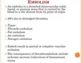
2.Propagation (progression) 3.Embolization 4.Organization and recanalization (inflammation and fibrosis)
If mass is large it undergoes • Organization: • the thrombus is invaded by capillaries and fibroblast • change to fibrous mass • lead to permanent vascular occlusion • Organization and Canalization; • some time capillaries dilated and allow Passage of blood through the thrombus;