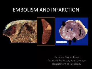
EMBOLISM AND INFARCTION
EMBOLISM AND INFARCTION. Dr Zahra Rashid Khan Assistant Professor, Haematology Department of Pathology. EMBOLUS. detached intravascular solid, liquid, or gaseous mass carried by the blood to a site distant from its point of origin causes partial/complete vascular occlusion.

EMBOLISM AND INFARCTION
E N D
Presentation Transcript
EMBOLISM AND INFARCTION Dr Zahra Rashid Khan Assistant Professor, Haematology Department of Pathology
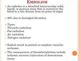
EMBOLUS • detached intravascular solid, liquid, or gaseous mass • carried by the blood to a site distant from its point of origin • causes partial/complete vascular occlusion
ORIGIN OF EMBOLI • 99% of all emboli arise form thrombus (hence the term thromboembolism) • OTHERS: fat droplets, bubbles of air or nitrogen, tumor fragments, bits of bone marrow, or foreign bodies
CONSEQUENCES • Pulmonarythromboembolism • Systemicthromboembolism
PULMONARY EMBOLISM • venous emboli originate from deep leg vein thrombi above the level of the knee • pass through the right side of the heart before entering the pulmonary vasculature • may occlude the main pulmonary artery, or pass out into the smaller, branching arterioles • frequently, there are multiple emboli
CLINICAL OUTCOMES Most pulmonary emboli (60% -80%) are clinically silent • sudden death, • right ventricular failure (corpulmonale) • pulmonary infarction • pulmonary hemorrhage *(dual blood supply of lung offsets infarction in some cases)
SYSTEMIC THROMBOEMBOLISM • emboli in the arterial circulation; usually result in infarction • (80%) arise from intracardiac mural thrombi
SYSTEMIC THROMBOEMBOLISM • in contrast to venous emboli, arterial emboli can travel to a wide variety of sites • common sites: lower extremities (75%) and the brain (10%) *paradoxical emboli
FAT EMBOLISM • Microscopic fat globules enter the circulation after fractures of long bones or after soft-tissue trauma
FAT EMBOLISM • Although fat and marrow embolism occurs in some 90% of individuals with severe skeletal injuries, only 10% patients have clinical findings • Fat embolism syndrome (pulmonary insufficiency, neurologic symptoms, anemia, and thrombocytopenia; fatal in about 10% of cases)
AIR EMBOLISM • air may enter the circulation during obstetric procedures or as a consequence of chest wall injury • more than 100 mL of air are required to produce a clinical effect • decompression sickness occurs when individuals are exposed to sudden changes in atmospheric pressure
AIR EMBOLISM Decompression Sickness • When air is breathed at high pressure, increased amounts of gas (particularly nitrogen) become dissolved in the blood and tissues • If the diver then ascends (depressurizes) too rapidly, the nitrogen expands in the tissues and bubbles out of solution in the blood to form gas emboli • Bends, Chokes, Caison’s disease
AMNIOTIC FLUID EMBOLISM • entry of amniotic fluid into maternal circulation via a tear in the placental membranes and rupture of uterine veins • grave but fortunately uncommon complication of labor and the immediate postpartum period • mortality rate of 20% - 40%
AMNIOTIC FLUID EMBOLISM • severe dyspnea,hypotensive shock, seizures and coma • ultimately, the patient develops pulmonary edema and DIC
INFARCTION • area of ischemic necrosis caused by occlusion of either the arterial supply or the venous drainage in a particular tissue • cardiovascular, cerebral, pulmonary, bowel infarction
INFARCTION • 99% of all infarcts result from thrombotic or embolic events, and almost all result from arterial occlusion • other mechanisms, such as local vasospasm,extrinsiccompression of a vessel (e.g., by tumor
INFARCTION: HISTOLOGY RED INFARCTS occur • with venous occlusion • in loose tissues • tissue with dual blood supply WHITE INFARCTS occur • with arterial occlusion • in solid organs
INFARCTION: HISTOLOGY • ischemic coagulative necrosis (inflammation followed by scar tissue) • brain is an exception • Septic infarctions occur when bacterial vegetations from a heart valve embolize or when microbes seed an area of necrotic tissue
FACTORS THAT INFLUENCE DEVELOPMENT OF AN INFARCT • Nature of blood supply (renal/splenicvsheaptic/pulmonary) • Rate of development of occlusion (collaterals/anastamosis) • Vulnerability to hypoxia (e.g, neurons) • Oxygen content of blood (anemia, cardiac disease)