ELECTRON TRANSPORT CHAIN/SYSTEM
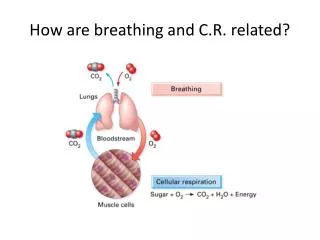
ELECTRON TRANSPORT CHAIN/SYSTEM. BIOT 309 Fall 2013. Electron transport chains are the cellular mechanisms used for extracting energy from sunlight in photosynthesis and also from redox reactions, such as the oxidation of sugars (respiration).

ELECTRON TRANSPORT CHAIN/SYSTEM
E N D
Presentation Transcript
ELECTRON TRANSPORT CHAIN/SYSTEM BIOT 309 Fall 2013
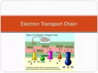
Electron transport chains are the cellular mechanisms used for extracting energy from sunlight in photosynthesis and also from redox reactions, such as the oxidation of sugars (respiration). • The electron transport chain consists of a spatially separated series of redox reactions in which electrons are transferred from a donor molecule to an acceptor molecule. The underlying force driving these reactions is the Gibbs free energy of the reactants and products. The Gibbs free energy is the energy available ("free") to do work. Any reaction that decreases the overall Gibbs free energy of a system is thermodynamically spontaneous.
Note that electrons can enter the chain at three levels: at the level of a dehydrogenase, at the level of the quinone pool, or at the level of a mobile cytochrome electron carrier. These levels correspond to successively more positive redox potentials, or to successively decreased potential differences relative to the terminal electron acceptor. In other words, they correspond to successively smaller Gibbs free energy changes for the overall redox reaction Donor → Acceptor.Individual bacteria use multiple electron transport chains, often simultaneously. Bacteria can use a number of different electron donors, a number of different dehydrogenases, a number of different oxidases and reductases, and a number of different electron acceptors. For example, E. coli (when growing aerobically using glucose as an energy source) uses two different NADH dehydrogenases and two different quinol oxidases, for a total of four different electron transport chains operating simultaneously.
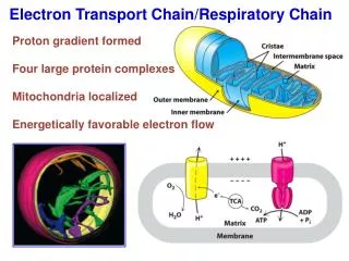
ELECTRON TRANSPORT CHAIN/SYSTEM • In bacterial membrane b/c • Allows charge separation • Stabilizes exact conformation • Responsible for: • Extruding protons and establishing proton motive force (pmf) across membrane (proton pump) • Removing electrons from substrates (NADH, FADH2, etc.) • Transferring electrons down a series of electron carriers to final electron acceptor, e.g., O2 during aerobic respiration • Not responsible for: generation of ATP; membrane bound ATP synthase does this
Although NADH is the most important electron donor in eukaryotes, bacteria can use a number of different electron donors, a number of different dehydrogenases, a number of different oxidases and reductases, and a number of different electron acceptors.
ETC COMPONENTS • Multi-subunit protein complexes join with electron carrier molecules • 1 or more electron carrier molecules associate with protein complexes depending on protein components • Depends on species, environmental conditions, even temperature
ETC COMPONENTS • Major classes of electron carrier types that undergo redox are: • Flavoproteins • Quinones • Iron-Sulfur proteins • Cytochromes
ETC/S GENERAL STRUCTURE • Dehydrogenase –> Quinones –> cytochromes (aerobic) • Dehydrogenase –> Quinones –> reductases (anaerobic) • Characteristics: Branched, modular, inducible • Multiple types of each => flexibility • Branches occur at quinones and cytochromes • Differ in Δ p generation b/c # coupling sites differ • Differ in final acceptor, e.g., oxygen, affinity • Bacterial ETC: species may use > 1 ETC b/c • More diverse • Genomes allow picking of system to suit environment, e.g., different carriers, different proteins • Different carbon sources • Different electron acceptors and donors
PRINCIPLE FOR ELECTRON MOVEMENT • E- move from high potential electron carriers to low potential electron carriers; each step involves redox reaction
ELECTRON TRANSPORT CHAINOF PROKARYOTES (AEROBIC)simple example
MORE COMPLEX ARRANGEMENT IN SOME BACTERIA • H+ moves outside membrane (proton motive force) • Electrons passed from one electron carrier to another • Not all extrude H+ Question 1: How many H+ move outside per ETC component? Question 2: Are these integral or peripheral proteins?
ETC OVERALL ATP PRODUCTION NADH + H+ + ½ O2+ 3ADP + 3 Pi —> NAD+ + 3ATP + 4 H20 NADH yields 3 ATP [FADH2 ] + ½ O2+ 2ADP + 2 Pi —> [FAD]+ + 2ATP + 3 H20 FADH2 yields 2ATP
FLAVOPROTEIN ELECTRON CARRIERS • Large enzyme complexes with flavins (FAD, FMN) as e- carrier • Named after electron donor, e.g., NADH dehydrogenase • Accept electrons from many sources: NADH, succinate • Pump protons (not in all bacteria) to periplasm • Funnel electrons to quinones • Associated with many different proteins -> different redox capabilities (potential) • Synthesized when needed, i.e., inducible REDOX
E. coli (glucose, aerobic) ETC • USES TWO • NADH dehydrogenases • different quinol oxidases • CARRIERS COMBINE INDEPENDENTLY • FOR A TOTAL OF FOUR DIFFERENT ETCS OPERATING SIMULTANEOUSLY
FLAVOPROTEINS STRUCTURES: RF, FMN, and FAD Adenine CH3 CH3 CH3 is terminal methyl group WHAT IS RIBOFLAVIN, other than a part of this molecule?
OXIDATION AND REDUCTIONFAD, FMN REDOX OCCURS AT N1 and N5 on riboflavin molecule
ETC: QUINONE ELECTRON CARRIERS • Mediators of electron transfer between different protein complexes in biological membranes • mobile carriers of electrons in membranes, but • some tightly associated with protein molecules to function in intramolecular electron transfer Example = NDH-1-type dehydrogenases (NDH-1) functioning as NADH:MK oxidoreductase • Lipids, lipid soluble • Can be highly mobile • Carry H+ and e- • side chains of varying lengths and differing saturation (-CH2-CH=C(CH3)-CH2-)n • Head groups can also vary • Ubiquinone and menaquinone • UQ (Coenzyme Q) – aerobic and nitrate respiration, mostly G- • MQ (structure related to vitamin K) – fumarate respiration, mostly G+, anaerobic G-, not in humans • Responsible for Q-cycle
OXIDIZED AND REDUCED FORMS REDOX Number of isoprenoid units( R) depends on species (-CH2-CH=C(CH3)-CH2-)n Head Group
QUINONE ELECTRON CARRIERS • Used by E. coli as terminal e- acceptor • E. coli does not have a cytochrome oxidase or a bc1 complex • Two different terminal quinol oxidases (both proton pumps) reduce oxygen to water
ETC: IRON-SULFUR PROTEIN ELECTRON CARRIERS • Non-heme iron and acid-labile sulfur - at biological pH the sulfur is bound to the iron • Non-heme iron bound to cysteine sulfur of a protein • Several iron sulfur clusters • Different Eh values • Proteins can have more than one Fe-S cluster • e- move around among them
ETC: IRON – SULFUR PROTEINS ELECTRON CARRIERS REDOX • Redox rxns in membrane (cytoplasm = non-ETC) • Non-heme iron bound to: • Sulfur of cysteine on protein (#) • “acid-labile” sulfur * (sulfide) * # * # LINEAR 2F - 2S CUBANE 4F - 4S SEVERAL DIFFERENT Fe-S CLUSTERS
ETC: IRON-SULFUR PROTEIN ELECTRON CARRIERS • Proteins can have more than one Fe-S cluster • Common bacterial clusters • F4-S4 • 2(F4-S4) • >2(F4-S4) • Variations at cys • Pyrococcus furiosus: Fe4S4, one of conserved Cys is substituted with aspartic acid • Loss of 4th cys also produces F3-S4
ETC: IRON-SULFUR PROTEIN ELECTRON CARRIERS • Protein structure determines if redox is: -1 -2 or -2 -3 (MW 6,000-9,0000) (Fe4S4*S4CYs)2- (Fe4S4*S4CYS)3- • Standard redox potentials do not apply b/c located within protein • Actual redox potential depends on protein • oxidoreductases
ETC: CYTOCHROME ELECTRON CARRIERS • Heme (non-heme Fe (NH-Fe)) = redox prosthetic group in proteins Fe 2+ Fe3+ • Have 4 pyrroles attached by methylene bridges • Pyrrole N linked with Fe • Different classes based on side chains of pyrrole rings • Different heme types in bacteria: a, b, c, d, o • Iron has redox capacity
TETRAPYRROLE/PORPHRYIN pyrrole methylene Tetrapyrrole or porphryin
ETC: CYTOCHROME ELECTRON CARRIERS • Different functions • Move 1 electron/heme • Most also act as proton pumps • Some are terminal oxidases and reductases in ETC, i.e., transfer to terminal acceptor EXAMPLE 4 Fe2+-cytochrome c + 8 H+in + O2 → 4 Fe3+-cytochrome c + 2 H2O + 4 H+out • Hemoproteins • Integral membrane proteins • Often have > 1 heme in a protein • Single or multiple subunits of protein • Entry point for electrons from inorganic e- donors: nitrite, ferrous ion, etc.
STRUCTURES: DIFFERENT HEMES * pyrrole * Heme b Cytochromes b Non-covalent bond to protein Heme a Cytochrome 3 oxidase Non-covalent bond to protein
STRUCTURES: DIFFERENT HEMES Heme c Cytochromes c Covalent bond to protein Heme o Cytochromes o Non-covalent bond to protein
DIFFERENT HEMES Heme d1 Occurs with cytochrome c -> cytochrome cd1 nitrate reductase Heme d Cytochromes d Non-covalent bond to protein
BACTERIA: DIFFERENT CYTOCHROMES C aa3 type: P. dentrificans, Rb. Sphaeroides b3 type: Thermus thermophilus bo3 type: E. coli ba3 type: Acetobacter aceti aa3 type: B. subtilis
TERMINAL ELECTRON ACCEPTORS • Inorganic and organic molecules • Inducible • Two general types • Aerobic uses OXIDASES: oxygen • Anaerobic uses REDUCTASES: fumarate, nitrate, nitrate
TERMINAL OXIDASE VARIATIONS • Cytochrome oxidases: a, aa3, b, d, o and combinations thereof, e.g., bd, cbb3 • Differ in: • affinities for oxygen • types of hemes or metals • ability to work as proton pumps • Quinol oxidases
ETC • Coupling sites: • Redox reactions coupled to H+ extrusion • Occur at dehydrogenase and oxidase complexes quinone shuttles electrons but does not perform redox
ETC in DIFFERENT BACTERIA • See White, Chapter 4.7 for ETC of different bacteria • See Figure 4.14, 4.15, 4.16 • E. coli is summarized next
SUMMARY: E. coli ETC • NADH dehydrogenase • NDH-1, coupling site; 4 H+/e- • NDH-2, not a coupling site no proton movement, only e- transfer • 3 quinones – ETC branches here • Type and % varies with e- acceptor(s) in use and [O2] • Aerobic: UQ = 60%, MQ = 3%, DMQ = 37% • Anaerobic: grow on nitrate – DMQ = 70%, MQ = 30% • Anaerobic: fumarate – MQ = 74%, DMQ = 16%, UQ = rest . ..
E. coli ETC • 2 quinol oxidases = terminal oxidases Aerobic characteristics • Quinol oxidation movement of 1 proton across membranes • cytochrome bd: = proton pump • b/c of Qox H+ moves outside • Operates at low O2 tension = high O2 affinity • cytochrome bo: = proton pump • 1H+/e- + 1 H+ from Qox • Operates at high O2 tension = low O2 affinity
EMP THROUGH ETC FLOW OF ELECTRONS AND ATP PRODUCTION