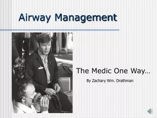
Airway management
Airway management. Dr. Rupak Bhattarai. Anatomy. There are two openings to the human airway, the Nose, which leads to the nasopharynx (pars nasalis), and the Mouth, which leads to the oropharynx (pars oralis).

Airway management
E N D
Presentation Transcript
Airway management Dr. Rupak Bhattarai
Anatomy • There are two openings to the human airway, the Nose, which leads to the nasopharynx (pars nasalis), and the Mouth, which leads to the oropharynx (pars oralis). • These passages are separated anteriorly by the palate, but they join posteriorly in the pharynx. • The pharynx is a U shaped fibromuscular structure that extends from the base of the skull to the cricoid cartilage at the entrance to the esophagus.
The pharynx opens anteriorly into the nasal cavity, the mouth, the larynx and the nasopharynx, and the laryngopharynx (pars laryngea) respectively. • The epiglottis prevents aspiration by covering the glottis –the opening of the larynx- during swallowing. • The larynx is a cartilaginous skeleton held together by ligaments and muscle. • The larynx is composed of nine cartilages thyroid, cricoid, epiglottic and in pairs arytenoid, corniculate and cuneiform.
THE EFFECTS OF LARYNGEAL NERVE INJURY ON THE VOICE • Superior laryngeal nerve: Unilateral :Minimal effects Bilateral: Hoarseness of voice • Recurrent laryngeal nerve Unilateral : Hoarseness of voice Bilateral : Acute : Stridor, Respiratory arrest Chronic: Aphonia • Vagus nerve Unilateral : Horseness of voice Bilateral: Aphonia
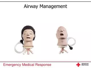
EQUIPMENT • ORAL AND NASAL AIRWAYS • Loss of upper airway muscle tone (eg. Weakness of the genioglossus muscle) in anesthetized patients allows the tongue and the epiglottis to fall back against the posterior wall of the pharynx. • Repositioning of the head or the insertion of the artificial airway is the preferred technique to create an air passage between the tongue and the posterior pharyngeal wall.
Types of oral airways • Guedel No (3): 80mm-(Small) • Guedel No (4): 90mm-(Medium) • Guedel No (5): 100mm-(Large) • The nasal airway are just 2-4cm longer than the oral airways.
Risk factors due to airways • Epistaxis in anticoagulated patients or in children with prominent adenoids. • Cough or even laryngospasm in awake or lightly anesthetized patients. • Injuries to the roof of the nasal passage and injuring to the vessels.
FACE MASK DESIGN AND TECHNIQUES • The use of facemask can facilitate delivery of oxygen or of an anesthetic gas from the breathing system to a patient by creating an airtight seal with the patient’s face. • TYPES OF FACEMASK • TRANSPARENT FACEMASK • BLACK RUBBER FACE MASK
Transparent masks allow observation of exhaled humidified gas and immediate recognition of vomiting. • Black rubber masks are pliable enough to adapt to uncommon facial structures (also called as anatomical face mask) • PARTS OF FACE MASK • THE RIM • THE ORIFICE • THE RETAINING HOOKS • THE BODY
PARTS OF FACE MASK • THE RIM • THE ORIFICE • THE RETAINING HOOKS • THE BODY
THE RIM: Attached to the variety of patient’s facial features. • THE ORIFICE : Attaches to the breathing circuit of the anesthesia machine. • THE RETAINING HOOKS : Can be attached to a head strap so that the mask does not have to be continually held in place with the hands. • THE BODY
TECHNIGUE OF HOLDING FACEMASK • The holding of face mask can be done by two ways: • ONE- HANDED FACE MASK TECHNIQUE • TWO-HANDED FACE MASK TECHNIQUE
ONE-HANDED FACE MASK TECHNIQUE • The mask is held against the face by downward pressure on the mask body exerted by the left thumb and index finger. • The middle and the ring finger grasp the mandible to facilitate extension of the atlanto-occipital joint. • Finger pressure should be placed on the bony mandible and not on the soft tissues supporting the base of the tongue , which may obstruct the airway. • The little finger is placed under the angle of the jaw and used to upthrust the jaw anteriorly , to allow ventilation to the patient.
TWO –HANDED FACE MASK TECHNIQUE • In difficult situations , two hands may be needed to provide adequate jaw thrust and create a mask seal. • Therefore an assistant may be needed to squeeze the anesthesia bag. • In such cases , the thumbs holds the mask down and the finger-tips displace the jaw forward. • CARE SHOULD BE TAKEN TO AVOID PRESSURE ON THE EYE, AND THE EYES SHOULD BE TAPED SHUT TO MINIMIZE THE RISK OF CORNEAL ABRASIONS.