Airway Management
1.32k likes | 2.75k Vues
Airway Management. Airway Physiology. Upper Airway. Begins at mouth and nose Air is warmed and humidified in nasal turbinates Jaw Throat / Pharynx Oropharynx Epiglottis Larynx/voice box Ends at glottic opening. Upper Airway. Lower Airway. Begins at glottic opening

Airway Management
E N D
Presentation Transcript
Upper Airway • Begins at mouth and nose • Air is warmed and humidified in nasal turbinates • Jaw • Throat / Pharynx • Oropharynx • Epiglottis • Larynx/voice box • Ends at glottic opening
Lower Airway • Begins at glottic opening • Trachea / Windpipe • Hollow tube which passes air to the lower airways • Supported by cartilage rings • Bronchi – branches at the carina • Lungs • Bronchioles • Thin hollow tubes that lead to alveoli • Remain open through smooth muscle tone • Alveoli
Alveoli • The end of the airway • Millions of tiny sacs in grapelike bunches at the end of the airway • Surrounded by capillary blood vessels • Oxygen and carbon dioxide diffuse through pulmonary capillary membranes
The Path of Oxygen • Oropharynx/Nasopharynx • Epiglottis • Larynx • Trachea – main tube that carries oxygen to the lungs • Right/Left Main Stem Bronchus • Bronchi • Alveoli
Purpose • Takes in oxygen • Disposes of wastes • Carbon dioxide • Excess water O2 + Glucose The Cell CO2 + H2O
Respiratory System Anatomy • Chest cage • Ribs • Muscles • Intercostal • Diaphragm • Pleura • Phrenic nerve innervation • Originates in C4 • Sends motor function to the diaphragm
Respiratory System Anatomy • Pleura • Double-walled membrane • Visceral layer covers lung • Parietal layer lines inside of chest wall, diaphragm
Respiratory System Anatomy • Diaphragm • Muscular structure that allows the body in inhale and exhale
Respiratory System Anatomy • Lung • Right lung 3 lobes • Left lung 2 lobes
Respiratory System Physiology • Pulmonary Ventilation • Ventilation is defined as the movement of air into and out of the lungs • Oxygenation • The amount of oxygen dissolved in blood and body fluids
Respiratory System Physiology • Respiration • Process by which the body captures and uses oxygen and disposes of carbon dioxide • External respiration • Exchange of oxygen and carbon dioxide between alveoli and the blood in the pulmonary capillaries • Internal respiration • Exchange of oxygen and carbon dioxide between the capillaries of the body tissues and the individual cells • Cellular respiration • Each cell of the body performs a specific function • Oxygen and sugar are essential to produce energy for cells to perform their function • Produce carbon dioxide as a waste product
Respiration Terminology • Tidal volume — amount of air moved in one breath • Dead space air — air moved in ventilation not reaching alveoli • Alveolar ventilation — air actually reaching alveoli • Ventilation — both inhaling and exhaling • Diffusion — movement of gases from high concentration to low concentration
Respiratory System Physiology • Inspiration • Active process • Chest cavity expands • Negative pressure pulls air into the lungs • Air flows in until pressure equalizes • Diaphragm lowers and contracts
Respiratory System Physiology • Expiration • Passive process • Muscles relax; size of chest decreases • Intrathoracic pressure rises • Air flows out until pressure equalizes • Diaphragm rises and relaxes
Respiratory System Physiology • Automatic Function • Primary drive: stimulus to breathe is based on high levels of arterial CO2 • Secondary (hypoxic) drive: stimulus to breathe is based on low levels of oxygen Normally we breathe to remove CO2 from the body, NOT to get oxygen in
Vascular Structures That Support Respiration • Pulmonary capillary structures • The heart • Right Heart • Receives system circulation • Drives pulmonary circulation • Left Heart • Receives pulmonary circulation • Drives system circulation • Arteries, arterioles, capillaries, venules, veins • Tissue/cellular beds
Airway Obstructions Tongue Foreign body airway obstruction Anaphylaxis-severe allergic reaction Upper airway burn Epiglottitis - children Croup - children Drowning Aspiration-crud goinginto the lungs Asthma Pneumonia Pulmonary edema Chronic Obstructive Airway Disease Emphysema Chronic bronchitis Respiratory Pathophysiology
Respiratory Failure Reduction of breathing to the point where oxygen intake is not sufficient to support life.
Respiratory Arrest Breathing stops completely.
Signs of Adequate Breathing • Look – bilateral chest expansion. Adequate & equal expansion on both sides. • Listen – Auscultate – to listen. Should be free of abnormal sounds (crackles or wheezing) • Feel – as the air is expelled from the nose and mouth. • Skin – normal color and tone
Pulse Oximetry • Assesses oxygenation • Quantify hemoglobin saturation • Complications (inaccurate readings) • shock patients • carbon monoxide poisoning • cold extremity
Normal Breathing Rates • Adult – 12-20 breaths per minute • Child – 15-30 breaths per minute • Infant – 25-50 breaths per minute Rhythm – regular rate, rhythm and quality Quality – breath sounds present and equal - chest expansion adequate Depth - adequate
Signs of Inadequate Breathing • Chest movements absent, minimal or uneven. • Abdominal breathing • Noises – wheezing, stridor, crackles, snoring respirations, silent chest. • Cyanosis – skin color is blueish/gray • Rate is too fast or too slow • Breathing is shallow, very deep and labored or irregular respiratory pattern
Signs of Inadequate Breathing 7.Inspirations are prolonged (possible upper respiratory obstruction). • Expirations are prolonged (possible lower airway obstruction). • Can not speak or can not speak in complete sentences. • Nasal flaring (infants & children) • Tripod Position – starving for oxygen • Changes in mental status - hypoxia
IF THE PATIENT DEMONSTRATES INADEQUATE VENTILATION (RESPIRATIONS OF LESS THAN 10 PER MINUTE OR GREATER THAN 29 PER MINUTE) AND THE PATIENT IS CONFUSED, RESTLESS, OR CYANOTIC THEN YOU MUST CONSIDER PROVIDING OXYGEN WITH A BAG-VALVE-MASK OR POCKET-MASK
A. Methods for Opening an Airway • Head-tilt, chin lift – used in a non-trauma patient. One hand should be placed on the forehead with the fingertips of the other hand under the lower jaw. • Jaw-thrust - used in a trauma patient where spinal precautions are a concern. Moves the mandible forward.
Pediatric Note forOpening the Airway • Infants and small children often have larger occipital regions of their heads • Lying flat may cause hyperflexion of neck and airway occlusion • Evaluate need to pad behind patient’s shoulders to achieve neutral airway position
B. Responsibilities of the EMT a. Be sure all equipment is clean and operating properly. b. Select proper equipment for patient care. c. Must monitor the patient closely. d. Must properly clean, discard and test all equipment after use.
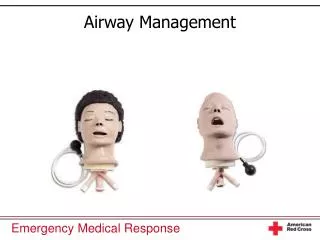
C. Airway Adjuncts • Most common airway obstruction is the tongue.
1. Oropharyngeal airway -ONLY use on an unconscious patient. -Device used to move tongue forward as it curves back to pharynx If there is a gag reflex, REMOVE IMMEDIATELY!!! - If the patient is unconscious assume a spinal cord injury based on the mechanism of injury. - Practice only on a manikin. - Can induce vomiting and/or bronchospasms. - Made in different sizes - you must measure for correct size. - Correct Size is the distance from the corner of the patient’s mouth to the tip of the earlobeon the same side of the face. (Alternate: measure from the center of the patient’s mouth to the angle of the lower jaw bone.)
Using Oral Airways If the patient becomes conscious, remove the airway and have suction ready. • Insert the airway upside down with the tip facing the roof of the mouth. • When resistance is encountered, turn the airway 180 degrees so that it comes to rest with the flange on the patient’s lips. • You may also insert the airway right side up, using a tongue depressor to press the tongue down. This is preferred in infants and children
2. Nasopharyngeal Airways - does not stimulate the gag reflex so it may be used on a patient with a reduced level of consciousness but still has an intact gag reflex. - can be used when teeth are clenched or patient has an oral injury - do not use if clear cerebrospinal fluid is coming from the ears or nose – look for the halo effect - made in different sizes - you must measure for correct size. - Correct Size is to measure from the tip of the nose to the earlobe or from the patient’s nostril to the angle of the jaw. Also, use the patient’s pinky finger to determine the diameter of the airway to be used.
Using Nasal Airways 1. Establish and maintain an open airway. 2. Lubricate with a water-based lubricant. 3. Insert into right nostril first, bevel of the airway toward the septum 4. If you feel resistance remove. Do not force! Try the other nostril. 5. Slide into the nose until the lip is against the nostril. 6. Remain ready to suction the patient if needed.
D. Suctioning Units A. Types - Oxygen or air powered - Electrically powered units (Current or battery) - Manual (bulb syringe)
Suction Systems • Fixed or portable
Suction Device Requirements • Must furnish air intake of at least 30 Lpm at open end of collection tube • Must generate vacuum of no less than 300 mmHg when collecting tube is clamped