Download
1 / 1
10 likes | 143 Vues
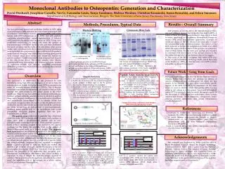
Future Work – Long Term Goals. Magnetic Bead Assay. ELISA. 0.25. 2. Native. 0.2. Native. Recombinant. 1.5. Recombinant. 0.15. 1. Optical Density (OD). Optical Density (OD). 0.1. 0.5. 0.05. 0. 0. 2A1. 10H4. 48F3. 49C12. 1. 2. 3. 4. 2A1. 2D6. 66G3. 45B5. 7E3. 71A10.

E N D
Future Work – Long Term Goals Magnetic Bead Assay ELISA 0.25 2 Native 0.2 Native Recombinant 1.5 Recombinant 0.15 1 Optical Density (OD) Optical Density (OD) 0.1 0.5 0.05 0 0 2A1 10H4 48F3 49C12 1 2 3 4 2A1 2D6 66G3 45B5 7E3 71A10 1 2 3 4 5 6 -0.5 -0.05 Antibodies Antibodies Monoclonal Antibodies to Osteopontin: Generation and CharacterizationDavid Denhardt, Josephine Cassella, Yao Li, Cassandra Louis, Tanya Gordonov, Melissa Weidner, Christian Kazanecki, Aaron Kowalski, and Esben SørensenDepartment of Cell Biology and Neuroscience, Rutgers, The State University of New Jersey, Piscataway, New Jersey Abstract Methods, Procedures, Typical Data Results – Overall Summary We have generated monoclonal antibodies (MAbs) to OPN using several strategies. OPN-deficient mice have been immunized with recombinant (unmodified) human and mouse OPN, with native human milk OPN (Christensen et al., 2005), and with peptides containing phosphorylated serines representative of selected, well-conserved phosphorylated sequences in OPN. Using well-established procedures, we fused cells from the spleens of immunized mice with SP2/0 myeloma cells and then distributed the fusion products into the wells of 96-well plates. After several weeks, cells from those wells that contained growing hybridomas were expanded into progressively larger cultures in rich medium; many died out along the way. Hybridomas showing good growth were weaned away from serum into serum-free hybridoma growth medium. Conditioned media were prepared and aliquots of the cells frozen down. The media samples were filtered, concentrated using Centripreps, and immunoglobulins purified on Sepharose Protein A/G columns. Anti-OPN monoclonal antibodies produced by these hybridomas are being characterized as described herein. Western Blotting Coomassie Blue Gels Our progress so far has led to the identification of many new and different monoclonal antibodies that bind to OPN. A subset of them have been characterized for their ability to bind to native (modified) or recombinant (unmodified) human or mouse OPN using four different approaches, examples of which are shown on the left. We have also been able to locate the epitopes that some of them recognize. The cartoon of the OPN molecule at bottom left (adapted from Sodek et al. 2000) shows the approximate location of the epitopes recognized by some of these MAbs. For the most part, epitope identification is consistent with the location of the epitope relative to the thrombin cleavage site. Our preliminary data so far suggest: 1) That some of the MAbs recognize only native OPN (e.g. 66A7), or only recombinant OPN (e.g. 73A3), and 2) that some of the MAbs (e.g. 45B5) are detected only when OPN is freely accessible, not bound to a support. A L S N/C thrombin fragments PC: polyclonal antiserum Native/recombinant OPN WB. →: Full length OPN Samples of concentrated, conditioned serum-free media are electrophoresed on SDS-PAGE gels and stained for protein using CB. L (~55kD) and S (~25kD) represent the approximate positions of the heavy and light IgG chains; A is an unknown protein. • Electrophorese proteins in an SDS-PAGE gel. • Transfer proteins in the gel onto a PVDF membrane. • Add antibodies to the strips of the membrane. • Add secondary antibody (Goat-anti-mouse HRP). • Identify MAb location by enhanced chemiluminescence. • Peptide Assay (similar to an ELISA) • Used to test which specific OPN sequence, if any, the monoclonal antibody binds. • Wells of 96-well neutravidin-coated plates are incubated with biotinylated OPN peptides. • 18-mer peptides (19 in all covering the entire OPN molecule) in separate wells. • The wells are washed and goat anti-mouse IgG conjugated to Alexafluor 594 is added. • After washing, the fluorescence (EX: 584 nm; EM: 590) is read. Readings above background suggest the MAb binds that peptide. Overview • Magnetic Bead Assay • Protein G coupled to magnetic beads (InVitrogen). • MAbs are incubated with the protein G/magnetic beads (protein G binds IgG at the Fc portion leaving the antigen binding site available). • Beads are washed, and differentially fluorescently labeled native and recombinant OPN are added. • The antibody/antigen complex is dissociated from the beads by low pH citrate buffer. • The fluorescence of the supernatant is read at the two wavelengths indicative of the 2 forms of OPN. Our goals in this research are two. On the one hand we want to identify MAbs that will allow the presence or absence of specific post-translational modifications, or exons for that matter, to be defined. This may allow functionally specific regions of the OPN molecule to be identified. On the other hand we want to identify MAbs that inhibit OPN function either in cell culture or in the animal. Functional inhibitors of native OPN in mammals might be useful therapeutic agents in the treatment of OPN-dependent pathologies; these include autoimmune disease, cancer metastasis, osteoporosis, stress-induced immune disfunction, and certain inflammatory conditions (sepsis?). Our approach is to characterize the IgG produced by each hybridoma using four different strategies so as not to bias the selection in favor of only MAbs that bound OPN in one particular assay, for example western blotting or ELISAs. This is because certain epitopes might be sequestered and unable to bind an MAb because they were bound to cell culture plastic or the PVDF membrane. Thus we use two additional screening methodologies to detect antigen-antibody associations free in solution (a peptide assay and a magnetic bead assay). We use both native (modified) and recombinant (unmodified) OPN to identify MAbs that specifically recognize either the native or the recombinant form. Antibodies specific only for the native form would be candidates for MAbs recognizing an epitope that is post-translationally modified; MAbs specific only for the recombinant form would presumably recognize peptide sequences that are post-translationally modified. The peptide assay makes use of peptides that collectively cover the entire OPN molecule with 2-4 amino acid overlaps; in some cases both a phosphorylated peptide and its non-phosphorylated counterpart are used. These peptides possess a biotin at the C terminus coupled to the peptide by two glycine residues; via the biotin, the peptides bind to the surface of NeutrAvidin-coated wells in a 96-well format. The magnetic bead assay employs two different forms of OPN, the highly post-translationally modified native OPN purified from human milk and the unmodified form synthesized in E. coli. In this novel assay the two forms (differentially labeled with fluorescent tags) are mixed and allowed to react in solution with a specific MAb. The antigen-antibody complexes are then captured on magnetic beads coupled to protein G. After the beads are washed, the adsorbed IgG is eluted at low pH, and the fluorescence at the two wavelengths determined. MAbs that selectively bind the native protein are presumed to be antibodies that recognize post-translational modifications. We believe this novel method is important in that it detects the antibody-antigen reaction in solution. This is in contrast to the methods currently in use which entail the immobilization of the antigen on a plastic surface (ELISA) or membrane (Western blots). Cartoon illustrating locations of know epitopes OPN molecule courtesy Jaro Sodek References Christensen B, Nielsen MS, Haselmann KF, Petersen TE, Sorensen ES. (2005) Post-translationally modified residues of native human osteopontin are located in clusters: identification of 36 phosphorylation and five O-glycosylation sites and their biological implications. Biochem J. 390:285-92. Sodek J, Ganss B, McKee MD. (2000) Osteopontin. Crit Rev Oral Biol Med. 11:279-303. Magnetic beads coupled to Protein-G . Acknowledgements This research was supported by funds from the NIH, a Busch Biomedical Research Award, the Rutgers Technology Commercialization Fund, and a grant from the National Multiple Sclerosis Society. The support of Aresty Summer Fellowships to Tanya Gordonov and Cassandra Louis are gratefully acknowledged. Yao Li was supported by a SURF award. We thank Avni Shah for technical assistance and Dr. Larry Steinman for his generous contributions of the peptides and other reagents used in this research. 2A1 is a positive control for native and recombinant OPN. 2D6 appears to bind both forms strongly; 66G3 binds more to native; 7E3 binds more to recombinant; 71A10 binds to neither; 45B5 shows a strong affinity for native OPN only in the magnetic bead and peptide assay; it has always appeared negative on Westerns and ELISAs. (IgG background subtracted.) 2A1 is a positive control for native and recombinant OPN. 10H4 also binds strongly to both forms, while 48F3 binds preferentially to native OPN whereas 49C12 binds more strongly to recombinant OPN.