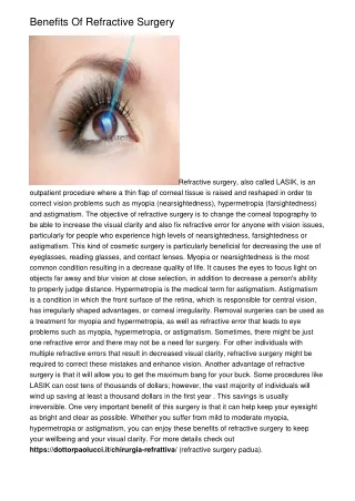
Quality Control in Refractive Surgery
Quality Control in Refractive Surgery. Stefan Pieger*, M.Sc. Wendelstein, Germany * Ingenieurbüro Pieger GmbH Nidek Germany Office. Introduction. Personal experience as application specialist for refractive excimer lasers since 1987. (Meditec, Schwind, Nidek)


Quality Control in Refractive Surgery
E N D
Presentation Transcript
Quality Control in Refractive Surgery Stefan Pieger*, M.Sc. Wendelstein, Germany * Ingenieurbüro Pieger GmbH Nidek Germany Office
Introduction • Personal experience as application specialist for refractive excimer lasers since 1987. (Meditec, Schwind, Nidek) • Progress in PRK&LASIK was usually based on outcomes analysis. • Excimer Laser Surgery and refractive surgery in general well suited for a systematic approach on quality control.
Why Quality Control? • Increase confidence level about refractive procedures offered in your center. • Verify current nomogram settings and make adjustments if necessary. • Reduce enhancement rate. • Use for marketing and advertisement. • Discover trends and technical problems in order to react more rapidly. • Fulfill requirements of ophthalmic societies.
How to collect your data? • Patients files • Excel Spread Sheet • Database Software (Access; Filemaker; etc.) • Outcomes Analysis Software (Datagraph; ASSORT; Refr. Consultant; etc.)
How to analyze refractive data? • Standard Refractive Outcomes • Stability / Safety / Predictability / Efficacy • Additional Outcome Parameters • Astigmatism Outcomes: Surgically Induced change in Cylinder (SIA); Double Angle Scatter Plot • Defocus Equivalent / Contrast Sensitivity in mesopic conditions / pre OP BSCVA vs. post OP UCVA • Wavefront Based Outcomes • Defocus + Cyl (‘aberrometer refraction’) • Higher Order RMS / Spherical Aberration (Z12) / Coma / Trefoil
Standard Refractive Outcomes: Safety 2 or more lines lost 3 % at 1 m post OP Number of eyes per Follow up visit. Follow up time interval
Standard Refractive Outcomes: Efficacy 72% 20/20 or better 3 month post Op Number of eyes per Follow up visit. Follow up time interval
Standard Refractive Outcomes: Stability (SEQ) ± 1 StDev Follow up time interval Mean value of SEQ Number of eyes per Follow up visit.
Standard Refractive Outcomes: Predictability (SEQ) Number of eyes at follow up ±1 D ‘happiness’ Zone Trend line y = -0.00x2 + 0.94x + 0.21 Regression Formula: ‘Achieved = 0.94*Attempted’ (~6% undercorrection)
Astigmatism Outcomes:SIA (based on Vector Analysis) -2/-1@180° 0/-0.5@90° (-0.5/+0.5@180°) 0.5 D Cyl Overcorrection! 0/-0.5@180° 0.5 D Cyl Undercorrection! y = 0.90 * x (~10% undercorrection)
Astigmatism Outcomes:Double Angle Scatter Plot PreOP Cyl & Axis PostOP Cyl & Axis
Additional Outcomes:Defocus Equivalent SEQ = SPH + ½ CYL DEQ = |SEQ|+|½ CYL|
Defocus Equivalent vs. Refractive Outcome -5.0/+10.0@90° SEQ Plano DEQ +5 DEQ ~ ‚Blur Circle‘
Additional Outcomes:Mesopic Contrast Sensitivity Normal Population Range (Vector Vision CSV 1000) VA in LogMar Scale
Wavefront based Outcomes – 2ndOrder / Coma / Trefoil • „Work in progress“ ! • Using Aberrometer Refraction rather than Manifest Refraction? • Presenting horizontal and vertical Coma individually? Vector calculation to present magnitude and axis in [D]! (0.5 D Coma @ 230°) • Trefoil: Axis? Present only magnitude?
Wavefront based Outcomes – Higher Order RMS [µm ±StDev] Pupil Diameter: 6.0 mm
Wavefront based Outcomes – Spherical Aberration [µm] or [D] Pupil Diameter: 6.0 mm
Making Outcome-based Nomogram Adjustments • Comparison of Laser Settings vs. Achieved change in refraction (and not Attempted vs. Achieved). • Reduce random errors as far as possible as nomograms can only compensate systematic errors! • Must be specific for major laser parameters like OZ, TZ, ablation profile type as well as for refraction types.
Nomograms for Individual Patient Groups • Data must be filtered on certain parameters: • Refraction Type (Myp/MyoAsti/Hyp/HypAsti…) • Surgery Type (PRK; LASIK; LASEK; Custom…) • Optical Zone Diameter • Others (age, laser software version; humidity…)
Nomogram Improvements – Laser Settings vs. Achieved y = -0,01x2 + 1,22x - 0,08 reduce attempted SEQ by 22%!
Laser Setting CYL vs. Surgical Induced change in Astigmatism 1. High Scatter! (further analysis necessary!) 2. 40 % systematic undercorrection
Nomograms: General Comments • Reduce Scatter by Standardized Surgery and OR Environment • Exclude Outliers from Data Analysis • Exclude Enhancements • Choose appropriate follow up interval (≥ 3 m) • Create Formula („-10%“) / Lookup Table or use Nomogram Software
Summary • Improving the results of refractive surgery procedures must be based on an individual quality control system. • Nomograms can compensate for systematic errors, but not for random errors. • Modern outcomes analysis software allows constant monitoring of your results. • Conventional Outcomes will be extended by HO-RMS, Spherical Aberration and Coma.
Thank You! www.datagraph-med.com