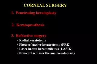
CORNEAL REFRACTIVE SURGERY
CORNEAL REFRACTIVE SURGERY. Major m kashif hanif DOMS.FCPS Cl. eye splt AFIO RWP. Sequence of presentation. Brief overview of anatomy of cornea Brief overview of physiology of cornea Corneal refractive surgery. Gross anatomy of cornea. 11.5mm horizontal diameter


CORNEAL REFRACTIVE SURGERY
E N D
Presentation Transcript
CORNEAL REFRACTIVE SURGERY Major m kashifhanif DOMS.FCPS Cl. eye splt AFIO RWP
Sequence of presentation • Brief overview of anatomy of cornea • Brief overview of physiology of cornea • Corneal refractive surgery
Gross anatomy of cornea • 11.5mm horizontal diameter • 10.5mm vertical diameter • 1 mm thick periphery • 0.5mm thick centrally • Anterior surface radius 7.7mm • Posterior surface radius 6.8mm
Microscopic anatomy • Epithelium • 5-6 layers thick • Stratified squamous , nonkeratinised • Superficial cells have microvilli • Basement membrane strongly attached to Bowman’s layer
Bowman’s layer • 8-12 microns • Acellular • interwoven collagen fibrils • Incapable of regeneration • Ends abruptly at limbus
Stroma • 90% of cornea thickness • 400microns centrally • 80% water • Glycosaminoglycans
Descemet’s membrane • 10microns thick • Type IV collagen fibrils • Basement membrane of the endothelium • Secreted and regenerated by endothelial cells • Terminates abruptly at limbus (Schwalbes line)
Endothelium • Single layer, polygonal, cuboidal cells • Tight junctions • Incapable of regeneration • Lines passages of trabecular meshwork
Physiology of cornea 3 main functions • Light transmission • Light refraction • Protection Corneal metabolism • Energy needed for maintenance of transparency and dehydration • Glucose • Oxygen
Ametropia • A refractive error (ametropia) is a disorder that occurs when parallel rays of light entering the non-accommodating eye are not focused on the retina
Normal Eye Myopia Astigmatism Hypermetropia
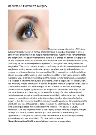
Aim Of Refractive Surgery • Alter refractive state of eye , enable patients to see without visual aids
refractive surgery divided into four major areas: • 1. INCISIONAL TECHNIQUES • 2. INTRASTROMAL CORNEAL RINGS (INTACS) • 3. THERMAL TECHNIQUES • 4. LAMELLAR PROCEDURES
Radial Keratotomy • Sato,1939 • radial cuts • pattern :spokes of a bicycle wheel • 4-16 incisions • Extra-pupillary region
Complications • Glare, star bursts • Fluctuation of vision • Regression, progression of refractive effect • Corneal perforation into the anterior chamber • Infectious keratitis and endophthalmitis
Incisional Astigmatic Keratotomy • Incision in the cornea flattens the meridian in which it is made and steepens the meridian 90 degrees away. • Single or paired • Optical zone between 6.0 mm and 7.0 mm
Relaxing incisions may be combined with compression sutures placed 90° away from them to reduce large degrees of corneal astigmatism
Intrastromal Corneal Rings • The ring segments flatten cornea similarly to the way you can flatten the top of a tent by pushing on the sides.
Intrastromal corneal Rings • Indicated for low myopia (1-3D) and min astigmatism < 1.00D • Advantage: reversible, natural corneal physiology maintained, no use of lasers , no removal of tissue
Complications • Fluctuation of vision • Under- or over-correction • Induced regular or irregular astigmatism • Glare , haloes • Corneal perforation • Pain • Infectious keratitis
Thermokeratoplasty • Hypermetropia +0.75 D to +3.25 D • Astigmatism less than or equal to 0.75 D • Spherical equivalent +0.75 D to +3.00 D
This technique modifies the central corneal curvature by heat-induced shrinkage of collagen fibers in the midperiphery of the cornea. • Noncontact Technique • Holmium laser thermokeratoplasty (LTK) • Conductive Keratoplasty • contact probe
Lamellar Procedures • Laser in situ keratomileusis • Laser Epithelial Keratomileusis • Epithelial LASIK
What is Laser? Light Amplification by Stimulated Emission of Radiation(LASER) • Ground state • Pumping • Atoms in the excited state are unstable and their electrons tend to spontaneously return to the ground state by emitting light energy
What Is An Excimer Laser? • EXCIMER= “excited dimer” • Argon or Xenon bound with a halogen eg. Fluorine or Chloride • diatomic gas halide - temporary excited state
Excimer Laser • During decay emits UV of 193nm • Remove controlled amounts of tissue with extreme precision
Photorefractive keratectomy • Indication • myopia up to 6D • astigmatism up to 3D • Hypermetropia up to 3D • Myopia • 10um of ablation = 1D of myopia • Hypermetropia
Photorefractive Keratectomy Technique • The visual axis is marked and the corneal epithelium removed • The patient fixates on the aiming beam of the laser • The laser is applied to ablate only bowmans layer and anterior stroma
30-60 seconds • Bandage contact lens • The cornea usually heals within 48-72 hours • Subepithelial haze • Post-op period • Topical steroids and NSAIDs. • Antibiotics
Complications • Slow healing epithelial defects • Corneal haze • Haloes • Dry eyes • Decentred ablations • Scarring • Abnormal epithelial healing • Irregular astigmatism • Hypoaesthesia • Sterile infiltrates • Infection
Laser EpitelialKeratomileusis • Indication • Myopia 8D • Hypermetropia 3D • Astigmatism 3D • LASEK works well for patients who are unsuitable for LASIK such as those with very thin cornea
Alchol 20% is applied for 30-40 seconds and an epithelial sheet is cleaved at the basement membrane • Laser is applied
Comlpications • Pain • Corneal haze • Over / undercorrection • Loss of sharpness of vision • Infection
Epithelial Lasik • Instead of using alcohol to loosen the epithelium, an epikeratome is used • Instead of using an oscillating sharp blade to incise the cornea beneath Bowman’s membrane, the epikeratome uses a blunt oscillating separator
LASER ASSISTED IN-SITU KERATOMILEUSIS (LASIK) • Concept first intro by Jose Barraquer, 1964 • founder : Dr IonasPallikaris- first to use microkeratome and laser • KERATOMILEUSIS = “to shape cornea” • IN-SITU = “ in place” • LASIK = “to shape cornea in place”
Inclusion Criteria • Myopia : 1-15D • Hypermetropia: 1-4D • Astigmatism : up to 5D • Adequate corneal thickness> 45O m
Inclusion Criteria • Age: > 21 years old, no max age. • stable refraction over past 24 months • Central K’s: > 39D - < 47D
Exclusion Criteria • Immunocompromise • Corneal irregularities • Pachymetry < 450m • Ocular herpes within the last year • Progressive myopia
Ideal Candidate • Be at least 18 years of age • Stable vision for at least one year • Adequate thickness of the cornea • Healthy eyes