Download
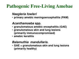
1 / 77
820 likes | 1.16k Vues
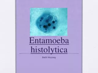
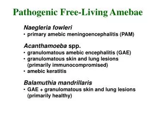
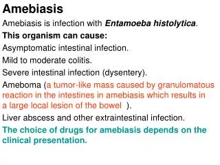
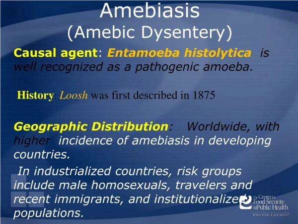
Amebiasis (Amebic Dysentery). History : Loosh was first described in 1875. Causal agent : Entamoeba histolytica is well recognized as a pathogenic amoeba. Geographic Distribution : Worldwide, with higher incidence of amebiasis in developing countries.

E N D
Amebiasis(Amebic Dysentery) History:Loosh was first described in 1875 Causal agent: Entamoeba histolytica is well recognized as a pathogenic amoeba. Geographic Distribution: Worldwide, with higher incidence of amebiasis in developing countries. In industrialized countries, risk groups include male homosexuals, travelers and recent immigrants, and institutionalized populations.
Morphology • Different form of E. histolytica; • 1- trophozoite • 2- precyst • 3- cyst(1, 2, 4 nuclei)
Trophozoite chractere • Size: 12-60μm in diameter; • Non-invasive form ( minuta) / E. dispare • Invasive form (magna) contain RBC, E. histolytica • Pseudopodia: • Motility: Ectoplasm: • Endoplasm: may be contain ingested RBC • Nucleoplasm: invasive form Non-invasive form
Life cycle Life cycle
Epidemiology • Prevalence of amebic infection varies with level of sanitation and generally higher in tropics and subtropics than in tempearate climates. • *Worldwide prevalence is about 10% to 50% • *Cyst passers are important source of infection • The true estimated prevalence of E. histolytica is close to 1% worldwide. • Entamoeba histolytica is the second leading cause of mortality due to parasitic disease in humans. (The first being malaria). Amebiasis is the cause of an estimated 50,000-100,000 deaths each year.
Transmission • 1-driect contact of person to person( fecal-oral) • 2- Veneral transmission among homosexual males( oral-anal • 3- Food or drink contaminated with feces containing the E.his. cyst • 4- Use of human feces (night soil) for soil fertilizer • 5- contamination of foodstuffs by flies, and possibly cockroaches
Pathogenesis • Effective factores: • 1- strain virulence: - classic strain - non-classic strain; Laredo , Huff, …. - pathogen zymodemes • 2- susceptibility of the host; nutrition status, immune-sys. • 3- breakdown of immunologic barrier (tissue invasion)
Pathogenicity mechanisms • 1- secreting proteolytic enzymes( histolysine ) and cytotoxic substances. • 2 - contact-dependent cell killing • 3 – cytophagocytosis • Amebic killing target cell: • 1- receptore-mediated adherence of amebae to target cell ( adherence lectin) • 2- amebic cytolysis of target cell • 3- amebic phagocytosis of killed target cell
Clinical symptoms Asymptomatic infection Symptomatic infection Intestinal Amebiasis Extraintestinal Amebiasis Dysenteric Non-Dysenteric colitisHepatic Pulmonary The extra foci Liver abscces Acut nonsupprative Intestinal Amebiasis symptoms: Diarrhea or dysentery, abdominal pain, cramping , anorexia, weight loss, chronic fatigue
Diagnosis • Paraclinical Diagnosis: • Sigmoidoscopic examination: precence of a grossly normal mucosa between the ulcers serves to differentiate amebic from bacillary dysentery,( the entire mucosa being involvoed in bacillary dysentery). • Hepatomegally • C.B.C. : leukocytosis in Amebic dys. rises above 12000 per microliter, but counts may reach 16000 to 20000 per microliter.
Laboratory Diagnosis • Entamoeba histolyticamust be differentiated from other intestinal protozoa including: E. coli, E. hartmanni, E. dispare,…… • Differentiation is possible, but not always easy, based on morphologic characteristics of the cysts and trophozoites. • The nonpathogenicEntamoeba dispar, however, is morphologically identical toE. histolytica, and differentiation must be based onisoenzymatic or immunologic analysis. • Molecular methods are also useful in distinguishing between E. histolytica and E. dispar and can also be used to identify E. polecki.
Microscopy • Microscopic identification This can be accomplished using: • Fresh stool: wet mounts and permanently stained preparations (e.g., trichrome). • Concentrates from fresh stool: wet mounts, with or without iodine stain, and permanently stained preparations (e.g., trichrome).
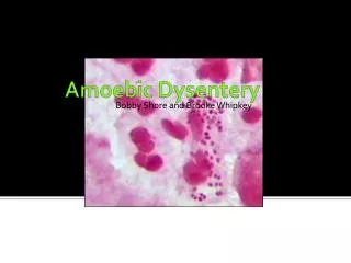
Trophozoites ofEntamoeba histolytica /E. dispar (trichrome stain) A B Microscopy In the absence of erythrophagocytosis, the pathogenic E. histolytica is morphologically indistinguishable from the nonpathogenic E. dispar! Each trophozoite has a single nucleus, which has a centrally placed karyosome and uniformly distributed peripheral chromatin.
Trophozoites ofEntamoeba histolyticawith ingested erythrocytes (trichrome stain) The ingested erythrocytes appear as dark inclusions. Erythrophagocytosis is the only morphologic characteristic that can be used to differentiateE. histolyticafrom the nonpathogenicE. dispar.
Cysts ofEntamoeba histolytica /E. dispar • GHI Cysts ofEntamoeba histolytica/E. dispar, permanent preparations stained with trichrome.
Immunodiagnosis(Antibody Detection) • 1- Antibody detection • 2- Antigen detection may be useful as an adjunct to microscopic diagnosis • The indirect hemagglutination (IHA) • The EIA test detects antibody specific for E. histolytica in approximately 95% of patients with extraintestinal amebiasis, 70% of patients with active intestinal infection, and 10% of asymptomatic persons who are passing cysts of E. histolytica.
Antigen Detection Antigen detection may be useful as an adjunct to microscopic diagnosis in detecting parasites and to distinguish between pathogenic and nonpathogenic infections. Recent studies indicate improved sensitivity and specificity of fecal antigen assays with the use of monoclonal antibodies which can distinguish betweenE. histolyticaandE. disparinfections.
Molecular diagnosis • In reference diagnosis laboratories, PCR is the method of choice for discriminating between the pathogenic species(E. histolytica)from the (nonpathogenic species (E. dispar.
Treatment • Intestinal Amebiasis: • *Asymptomatic amebiasis(cyst passer): Diloxanide furoate ( furamide) 500 mg 3 times daily / 10 days • *Symptomatic amebiasis ( troph. & cyst): - Iodoquinol , 650 mg 3 times daily/ 20 days or Metronidazole (Flagyl) , 750 mg 3 times daily/ 10 days • *Amebic colitis: Chloroquine, 250 mg 2 times daily • * Acute amebic dysentery: Emetine hydrochloride, 1mg/kg daily IM or SC
Treatment • Extraintestinal Amebiasis: • *Amebic liver abscess, ameboma: Metronidazole, as above plus dehydroemetine / 10 days or Metronidazole or dehydroemetine as above plus Chloroquine , 500 mg 2 times daily / 2 days,…..
Overview • Organism • History • Epidemiology • Transmission • Disease in Humans • Disease in Animals • Prevention and Control Center for Food Security and Public Health, Iowa State University, 2013
Organism • Giardia intestinalis • Protozoal parasite • Also known as: • Giardia lamblia • Lamblia intestinalis • Giardia duodenalis • Isolated from humans, domestic animals, and wild animals Center for Food Security and Public Health, Iowa State University, 2013
Organism • Human infections • Humans are main reservoir • Interspecies/zoonotic transmission • Importance of animal reservoirs unclear • Non-zoonotic Giardia spp. found in: • Rodents • Birds • Reptiles • Amphibians Center for Food Security and Public Health, Iowa State University, 2013
History • 1681 • van Leeuwenhoek, the “Father of Microbiology,” observes Giardia trophozoites in his own stool • Doubt common regarding pathogenicity of Giardia organisms • 1970s • Symptomatic travelers from Soviet Union increased awareness Center for Food Security and Public Health, Iowa State University, 2013
Geographic Distribution • Giardia intestinalis • Occurs worldwide • Most common in warm climates Center for Food Security and Public Health, Iowa State University, 2013
Morbidity and Mortality: Humans • Populations affected • Children • Travelers, hikers • Swimmers • Prevalence in developed countries • 2% of adults • 6-8% of children • Up to 15% in developing countries Center for Food Security and Public Health, Iowa State University, 2013
Morbidity and Mortality: Humans • Naïve populations • Morbidity rate up to 20% • Infections often resolve spontaneously • Chronic infections occur • May contribute to decreased lifespan in immunodeficient individuals Center for Food Security and Public Health, Iowa State University, 2013
Morbidity and Mortality: Animals • Young animals most affected • Reported prevalence rates • Puppies: 20-35% • Kittens: 10-15% • Foals: 17-32% • Calves: 5-90% • Lambs: 6-80% • Pigs: 7-44% • Usually not life threatening Center for Food Security and Public Health, Iowa State University, 2013
Giardiasis Incidence, 2011 Center for Food Security and Public Health, Iowa State University, 2013
Giardia Case Reports, by Age, 2006-2008 Center for Food Security and Public Health, Iowa State University, 2013
Parasite Stages • Two stages of the parasite: cyst and trophozoite Center for Food Security and Public Health, Iowa State University, 2013
Transmission • Cysts • Direct transmission • Fomites • Contaminated water and/or food • Ingested cysts release trophozoites • Trophozoites multiply and encyst in intestines • Excreted in feces Center for Food Security and Public Health, Iowa State University, 2013
Survival • Cysts • Survive well in cool, moist conditions • Remain viable for months in cold water • Two months at 8oC • One month at 21oC • Can also survive freezing • Susceptible to desiccation and direct sunlight Center for Food Security and Public Health, Iowa State University, 2013