Amebiasis
Amebiasis . Amebiasis is a common intestinal protozoal infection that may also cause systemic manifestations. About 90% of infections are asymptomatic, and the remaining 10% produce a spectrum of clinical syndromes ranging from dysentery to abscesses of the liver or other organs. Background.

Amebiasis
E N D
Presentation Transcript
Amebiasis is a common intestinal protozoal infection that may also cause systemic manifestations. About 90% of infections are asymptomatic, and the remaining 10% produce a spectrum of clinical syndromes ranging from dysentery to abscesses of the liver or other organs.
Background • Amebiasis is a parasitic infection caused by the protozoon Entamoeba histolytica. • Amebiasis is the third leading parasitic cause of death worldwide.
Life cycle Infection by Entamoeba histolytica 1 . Occurs by ingestion of mature cysts in fecally contaminated food, water, or hands. 2 . Excystation occurs in the small intestine and 3 . Trophozoites are released, which migrate to the large intestine. 4 . Trophozoites multiply by binary fission and produce cysts , which are passed in the feces.Because of the protection conferred by their walls, the cysts can survive days to weeks in the external environment and are responsible for transmission. (Trophozoites can also be passed in diarrheal stools, but are rapidly destroyed once outside the body, and if ingested would not survive exposure to the gastric environment.)
. • In many cases, the trophozoites remain confined to the intestinal lumen ( A: non-invasive infection) of individuals who are asymptomatic carriers, passing cysts in their stool. • In some patients the trophozoites invade the intestinal mucosa ( B : intestinal disease), or, • Pass through the bloodstream, extraintestinal sites such as the liver, brain, and lungs (C : extra-intestinal disease), with resultant pathologic manifestations. The invasive and noninvasive forms represent two separate species, respectively E. histolytica and E. dispar, however not all persons infected with E. histolytica will have invasive disease. These two species are morphologically indistinguishable.
. The parasite has 2 forms: - a motile form, called the trophozoite, and - a cyst form - The trophozoite of E histolyticainhabits the large intestine to produce lesions of amebic colitis. • Invasion of the colonic mucosa leads to dissemination of the organism to extracolonic sites, predominantly the liver.
. • Cysts passed in the feces can survive in moist environmental conditions for weeks to months. Upon ingestion of fecally contaminated food or water, the cysts travel to the small intestine, where the trophozoites are released. • In 90% of patients, the trophozoites re-encyst and produce asymptomatic infection, which usually spontaneously resolves within 12 months. • In the remaining 10% of patients who are infected, the parasite causes symptomatic amebiasis. • Under unfavorable conditions, the trophozoite reverts to the cyst form, and the life cycle is repeated.
. • Entamoeba dispar is a nonpathogenic protozoon morphologically identical to E histolytica. • These 2 species of Entameba can be distinguished by the monoclonal antibodies. • Specific and sensitive means to detect E histolytica in stool are now available and include antigen detection and polymerase chain reaction (PCR).
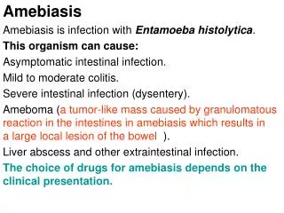
Infections due to E histolytica • Intestinal disease • Asymptomatic infection • Symptomatic noninvasive infection • Acute proctocolitis( inflammation of the rectum and colon ) • Fulminant colitis (coming on suddenly with great severity ) with perforation • Toxic megacolon (dialated colon ) • Chronic nondysenteric colitis • Ameboma (an inflamed, tumor like, spreading nodule ) • Perianal ulceration • Extraintestinal disease • Liver abscess • Pleuropulmonary disease • Peritonitis • Pericarditis • Brain abscess • Genitourinary disease
Pathophysiology • fecal-oral route • excystation in the small bowel and invasion of the colon by the trophozoites. • Invasive disease begins with the adherenceof E histolytica to colonic mucins, epithelial cells, and leukocytes. • Adherence of the trophozoite is mediated by a galactose-inhibitable adherence lectin.
Cont .. • After adherence, trophozoites :- • invade the colonic epithelium to produce the ulcerative lesions typical of intestinal amebiasis . • lyse the target cells by using lectin to bind to the target cells' membranes and using the parasite's ionophore like protein to induce a leak of ions (i.e, Na+, K+, Ca+) from the target cell cytoplasm. • An extracellular cysteine kinase causes proteolytic destruction of the tissue, producing flask-shaped ulcers
Cont .. • Spread of amebiasis to the liver occurs via the portal blood. • Trophozoites ascend the portal veins to produce liver abscesses filled with acellular proteinaceous debris. This material has the appearance of anchovy paste. • The trophozoites of E histolytica lyse the hepatocytes and the neutrophils. This explains the paucity (smallness of quantity ) of inflammatory cells within the liver abscesses.
Cont .. • Serum antibodies in patients with amebic liver abscess develop in 7 days and persist for as long as 10 years. • E dispar infections do not elicit antibody response • Mucosal immunoglobulin A (IgA) response to E histolytica occurs during invasive amebiasis. • However, no evidence suggests that invasive amebiasis is increased in incidence or severity in patients with IgA deficiency.
Cont .. • Cell-mediated immunity is important in limiting the disease and preventing recurrences. • production of lymphokines, including interferon-d (IFN-d), which activates the killing of E histolytica trophozoites by the macrophages. This killing depends on contact, oxidative pathways, nonoxidative pathways, and nitric oxide (NO). • Lymphokines, such as tumor necrosis factor-alpha (TNF-a), are capable of activating the amebicidal activity of neutrophils. • During acute invasive amebiasis, T-lymphocyte response to E histolytica antigens is depressed by a parasite-induced serum factor.
Mortality/Morbidity • Mortality rate in patients with uncomplicated amebic liver abscess is less than 1%. • Fulminant amebic colitis has a mortality rate of more than 50%. • Pleuropulmonary amebiasis has a mortality rate of 15-20%. • Amebic pericarditis has a case fatality rate of 40%. • Cerebral amebiasis is highly fatal, with a 90% death rate. Increased severity of amebiasis is noted : • in children (especially neonates), • women who are pregnant or postpartum, individuals who use corticosteroids, • individuals with malignancy, and • malnourished individuals.
Cont .. Sex • Invasive amebiasis, including amebic liver abscess, is much more common in adult males than in females. However, amebic liver abscess is equally common in both sexes among prepubertal children. Age • Symptomatic intestinal amebiasis occurs in all age groups. • Liver abscesses due to amebiasis are 10 times more frequent in adults than in children.
Diagnosis Clinical History • The incubation period is commonly 2-4 weeks but ranges from a few days to years. • Amebiasis is more severe in very young patients, in elderly patients, and in patients receiving corticosteroids. • The clinical spectrum of amebiasis ranges from asymptomatic infection to fulminant colitis and peritonitis to extraintestinal amebiasis, most commonly amebic liver abscess.
Cont .. • Asymptomatic infections are common following ingestion of the parasite. E dispar does not cause invasive disease or antibody production. • As many as 90% of E histolytica infections are also asymptomatic. The infection is self-limited but may be recurrent. • Only antigen detection tests can distinguish between E histolytica and E dispar.
Cont .. Amebic colitis : - • gradual in onset, with symptoms presenting over 1-2 weeks, • Diarrhea is the most common symptom. • cramping abdominal pain, watery or bloody diarrhea, and weight loss. • Fever is noted in 10% of patients. • Fulminant (coming on suddenly with great severity )amebic colitis : - • is a rare complication of amebic dysentery (<0.5%). • It presents with a rapid onset of severe bloody diarrhea, severe abdominal pain, and high fever. • Children younger than 2 years are at increased risk. • Intestinal perforation is common.
Cont .. • Patients may develop toxic megacolon, which is typically associated with the use of corticosteroids. • Chronic amebic colitis is clinically similar to inflammatory bowel disease. Recurrent episodes of bloody diarrhea and vague abdominal discomfort develop in 90% of patients with chronic amebic colitis who have antibodies to E histolytica. • Amebic colitis should be ruled out prior to treatment of suspected inflammatory bowel disease because corticosteroid therapy worsens amebiasis.
Cont.. • A less common form of intestinal disease, ameboma, • results from formation of annular colonic granulation in response to the infecting amebae, resulting in a large local lesion of the bowel. • It presents as a right lower quadrant abdominal mass, which may be mistaken for carcinoma,tuberculosis, Crohn`s disease, or lymphoma. • Biopsy findings assist in establishing the correct diagnosis.
Cont.. • Amebic liver abscess is the most common form of extraintestinal amebiasis. • It results from spread of the organisms from the intestinal submucosa to the liver via the portal system. • Approximately 40% of patients who have amebic liver abscess do not have a history of prior bowel symptoms. • 5% of patients with symptomatic intestinal amebiasis and is 10 times as frequent in men as in women.
Cont .. • presents with fever and a constant, dull, upper right abdominal or epigastrium pain. • Involvement of the diaphragmatic surface of the liver may lead to right-sided pleuritic pain or referred shoulder pain. Associated GI symptoms : - • occur in 10-35% of patients and include nausea, vomiting, abdominal distention, diarrhea, and constipation. • May present with vague abdominal discomfort, weight loss, and anemia.
Cont... • Pleuropulmonary amebiasis is most commonly the result of contiguous spread from a liver abscess rupturing through the right hemidiaphragm. • cough, pleuritic pain, and dyspnea. • Hepatobronchial fistula expectoration of sputum resembling anchovy paste. The trophozoites of E histolytica may be found in the sputum sample. • Amebic peritonitis is generally secondary to a ruptured liver abscess. Left lobe liver abscesses are more likely to rupture. Patients present with fever and rigid distended abdomen. • Roughly 2-7% of liver abscesses rupture into the peritoneum.
Cont.. Amebic pericarditis : - • caused by a rupture of the left liver lobe abscess and occurs in 3% of patients with hepatic amebiasis. It presents with chest pain and the features of congestive heart failure. Cerebral amebiasis : - • abrupt onset and rapid progression to death in 12-72 hours. • presents with altered consciousness and focal neurologic signs. • CT scanning reveals irregular lesions without a surrounding capsule. • Tissue biopsy :- trophozoites. Genitourinary involvement : - • may cause painful genital ulcers or fallopian tube amebiasis.
Physical Acute amebic colitis : - • lower quadrant abdominal tenderness. • Fever is noted in only a minority of patients. • Dehydration is uncommon. • Occult blood is nearly always present in stools. Amebic liver abscess : - - fever and tender hepatomegaly. - Right lower intercostal tenderness, particularly posteriorly. - Breath sounds may be diminished at the right lung base, and rales may be heard. - hepatomegaly, weight loss, and anemia. Pleuropulmonary amebiasis:- • produce findings of right-sided pleural effusions, empyema, pneumonia, and lung abscess.
Cont .. Amebic peritonitis : - • present with fever and a tender, rigid, and distended abdomen. Amebic pericarditis : - • presents with features of congestive heart faliure. • pericardial friction rub may be audible. Cerebral amebiasis : - presents with altered consciousness and focal neurologic signs. CT scanning reveals irregular lesions without a surrounding capsule or enhancement. Genital ulcers :- punched-out appearance and profuse discharge.
Differential Diagnosis • Intestinal amebiasis should be distinguished from the following conditions: • Infectious conditions • Campylobacter • Shigella • Salmonella • Yersinia • Enteroinvasive Escherichia coli • Enterohemorrhagic E coli
Cont.. • Noninfectious conditions • Inflammatory bowel disease • Diverticulitis Amebic liver abscess should be distinguished from the following conditions: • Pyogenic liver abscess • Necrotic hepatoma • Echinococcal cyst • The probability of a liver abscess to be amebic rather than pyogenic is increased by the history of residence in or recent travel to endemic areas, male sex, increased age (>50 y), presence of a single lesion in the right lobe of the liver, and the absence of jaundice, biliary disease
Laboratory Studies Stool Light microscopy: • Examination of a fresh stool smear for trophozoites that contain ingested RBCs. • Routine microscopy cannot distinguish the E dispar (nonpathogenic amebae) from E histolytica. • PCR-based diagnostic tests • Other stool tests • The stool samples are always heme positive. • Fecal leukocytes may be absent.
Serum tests • Antibody tests: • Indirect hemagglutination antibody (IHA) test • Detection of immunoglobulin M (IgM) antibodies • ELISA for Ab detection (most sensitive test) • Sigmoidoscopy or biopsy of symptomatic sick patients.
Imaging Studies Chest radiography : - • elevated right hemidiaphragm and a right-sided pleural effusion in patients with amebic liver abscess. • Ultrasonography (amebic liver abscess ) • cerebral amebiasis, CT shows irregular lesions without a surrounding capsule • MRI
Other Tests • Leukocytosis without eosinophilia is observed in 80% of cases. • Mild anemia may be noted. Liver function tests : - • elevated alkaline phosphatase levels (in 80% of patients), • elevated transaminase levels, • mild elevation of serum bilirubin level, • Erythrocyte sedimentation rate is elevated.
Treatment • Medical Care • Luminal agents that are minimally absorbed by the GI tract (eg, paromomycin, iodoquinol, diloxanidefuroate) are best suited for such therapy. • Metronidazole is the mainstay of therapy for invasive amebiasis. • Tinidazole for intestinal or extraintestinalamebiasis. • Other nitroimidazoles with longer half-lives (ie, secnidazole, ornidazole) • Nitroimidazole therapy leads to clinical response in approximately 90% of patients with mild-to-moderate amebic colitis. • Chloroquine has also been used for patients with hepatic amebiasis.
Cont.. • Intraluminal parasites are not affected by nitroimidazole therapy. Therefore, nitroimidazole therapy should be followed by treatment with a luminal agent such as paromomycin or diloxanide furoate to prevent a relapse. • Broad-spectrum antibiotics may be added to treat bacterial superinfection in a case of fulminant amebic colitis and suspected perforation.
Surgical Care Surgical intervention :- • perforated amebic colitis, massive GI bleeding, or toxic megacolon. • Amebic liver abscess generally responds to medical therapy alone and drainage is seldom necessary. • When necessary, imaging-guided percutaneous treatment (needle aspiration or catheter drainage) has replaced surgical intervention as the procedure of choice for reducing the size of an abscess. Indications for drainage : - • Presence of left-lobe abscess (>10 cm in diameter) • Rupture and • abscess that does not respond to medical therapy within 3-5 days
Antibiotics - Metronidazole is considered the drug of choice for symptomatic, invasive disease. • Paromomycin is the drug of choice for noninvasive disease. Because parasites persist in the intestine of 40-60% of patients treated with metronidazole, follow it with paromomycin to cure luminal infection. • Do not give the 2 medications at the same time because the diarrhea that often results from paromomycin might be confused with continuing active intestinal disease from the parasite.
Metronidazole (Flagyl, Protostat ) • Kills trophozoites of E histolytica in intestine and tissue. Does not eradicate cysts from intestines. • Adult • Intestinal amebiasis:PO: 500-750 mg PO tid for 5-10 d; alternatively, 2 g PO qd for 3 d or a single dose of 50 mg/kgIV: 500 mg IV q6h for 5-10 dAmebic liver abscess: 500 mg IV q6h for 10 d • Pediatric • 35-50 mg/kg/d PO/IV divided q8h for 10 d
Tinidazole (Fasigyn, Tindamax) • 5-nitroimidazole derivative with selective antimicrobial activity against anaerobic bacteria and protozoa. The mechanism by which tinidazole exhibits activity against Giardia and Entamoeba species is not known. • Adult • Intestinal amebiasis: 600 mg bid or 800 mg tid PO for 5 d; alternatively, 2 g PO qd for 3 d with foodHepatic amebic abscess: 2 g PO qd for 3-5 d with food • Pediatric • <3 years: Not established>3 years:Intestinal amebiasis: 50 mg/kg/d PO for 3 d with food; not to exceed 2 g/doseAmebic liver abscess: 50 mg/kg/d PO for 3-5 d with food; not to exceed 2 g/dose, limited data exist for
Paromomycin ( Humatin ) Amebicidal aminoglycoside antibiotic that is poorly absorbed. Active only against intraluminal form of amebiasis. Used to eradicate cysts of E histolytica following treatment with metronidazole or tinidazole for an invasive disease. • Adult • 25-35 mg/kg/d PO divided q8h for 7 d • Pediatric • Administer as in adults
Anthelmintics • Iodoquinol (Yodoxin) • Halogenated hydroxyquinoline. Luminal amebicide; acts primarily in bowel lumen because it is poorly absorbed. Best tolerated when given with meals. Because it is active only against intraluminal form of amebiasis, it is used to eradicate cysts of E histolytica after treatment of invasive disease. • Adult • 650 mg PO tid for 20 d • Pediatric • 30-40 mg/kg/d PO divided tid for 20 d; not to exceed 2 g/d
Chloroquine phosphate (Aralen) • Inhibits growth by concentrating within acid vesicles of parasite, which increases internal pH of organism. Also inhibits hemoglobin utilization and metabolism of parasite. In vitro studies with trophozoites of E histolytica demonstrate that chloroquine possesses amebicidal activity. • Highly effective in treatment of amebic liver abscess when administered with emetine or dehydroemetine. • Adult • Hepatic amebiasis:500 mg salt (300-mg base) PO bid for 2 d, followed by 250 mg salt (150-mg base) bid for 2-3 wk • Pediatric • Hepatic amebiasis: 10 mg (as base)/kg/d PO divided bid for 2-3 wk
Diloxanide furoate (Furamid, Entamizole, Furamide) • Luminal amebicide; acts primarily in bowel lumen because it is poorly absorbed. Used to eradicate cysts of E histolytica after treatment of invasive disease. • Adult • 500 mg PO tid for 10 d • Pediatric • 20 mg/kg/d PO divided tid for 10 d; not to exceed 1500 mg/d
Follow-up Prevention • Improved sanitation is critical to preventing orofecal transmission of organisms such as E histolytica. • Travelers to developing countries should be advised to avoid consumption of unsafe food and water. • Eating only cooked food or self-peeled fruits in endemic areas minimizes the risk. • Travelers should avoid eating raw fruits and salads, which are difficult to sterilize. • The amount of chlorine normally used to purify water is inadequate in killing the cysts. Drinking water can be rendered safe by boiling • Bottled water may be used for drinking when traveling to endemic areas.
Complications • Bowel perforation • GI bleeding • Stricture formation • Fistula formation • Intussusception • Secondary bacterial infection of amebic liver abscess (uncommon) • Peritonitis • Pericarditis • Empyema • Brain abscess
Prognosis • Intestinal infections due to amebiasis generally respond well to appropriate therapy. The severity of amebiasis is increased in the following individuals: • Children, especially neonates • Pregnant and postpartum women • Those using corticosteroids • Those with malignancies • Malnourished individuals • uncomplicated amebic liver abscess is less than 1%. • Fulminant amebic colitis has a mortality rate of more than 50%. • Pleuropulmonary amebiasis has a 15-20% mortality rate. • Amebic pericarditis has a case fatality rate of 40%. • Cerebral amebiasis is highly fatal, with a 90% death rate.
Patient Education • Educate patients about the prevention : - • avoiding drinking contaminated water and avoiding eating raw fruits and salads, which are difficult to sterilize. • Bottled water may be used during such travel. • Eating only cooked food in endemic areas minimizes risk.