Pediatric Cardiac Anomalies
260 likes | 571 Vues
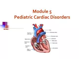
Pediatric Cardiac Anomalies. Congenital Heart Disease NPN 200. Fetal Cardiac Circulation. inferior vena cava to the right atrium then through the foramen ovale to the left atrium

Pediatric Cardiac Anomalies
E N D
Presentation Transcript
Pediatric Cardiac Anomalies Congenital Heart Disease NPN 200
Fetal Cardiac Circulation • inferior vena cava to the right atrium then through the foramen ovale to the left atrium • Superior vena cava to the right ventricle through the pulmonary artery to the ductus arteriosus and into the descending aorta • Very little blood goes to the lung • With 1st breath the pressure increases in the pulmonary vascular system and systemic pressure increases • The foramen ovale closes and the ductus arteriosus starts to close with the ^ O2 supply
Atrial Septal Defect • Abnormal flowing of blood between the atrium, allowing blood to flow from the left atria into the right atrium • Allows oxygenated blood in the left atrium to communicate with unoxygenated blood in the right atrium • May not cause any problems until later in life • May have atrial atrial dysrhythmias • May repair with Dacron graft
Ventricular Septal Defect • Abnormal opening between the right and left ventricle • May vary in size from a pin hole to absence of the septum • Frequently associated with pulmonary stenosis, transposition of the great vessels, patent ductus, atrial defects, and coarctation of the aorta • Many will close spontaneously, during the 1st year of life • Oxygenated blood flows into the lungs and increases pulmonary vascular resistance • The right ventricle hypertrophies due to the increase in pressure • Right atrial enlargement may occur
VSD, cont. • CHF is common • Murmur • Risk for bacterial endocarditis, and pulmonary vascular obstructive disease • May use band around the pulmonary to decrease blood flow into the lungs • May also use Dacron graft or purse string suture
Patent Ductus Arteriosus • Failure of ductus to close within the first few weeks of life • Murmur will be present • Bounding pulse • At risk for endocarditis and pulmonary vascular disease in later life • Treatment – • Indocin will help close in some premature babies • Surgical repair by opening the chest or now may use laparoscope
Pulmonary Stenosis/Aortic Stenosis • Aortic stenosis = narrowing or stricture of aortic valve • Pulmonary stenosis = narrowing of the entrance to the pulmonary artery • Both require interventions if severe enough to cause problems of CHF and heart failure • May be repaired by replacement or ballooning
Tetralogy of Fallot • 4 defects • Ventricular septal defect • Pulmonary stenosis • Overriding aorta – entrance of the aorta is close to the VSD • Right ventricular hypertrophy
Patho • Amount of problems depend upon the amount of pulmonary stenosis and the size of the VSD • Most common defect with causes cyanosis • Defects causes the blood flow to shunt from one chamber to another and pulmonary stenosis prevents the blood from going to the heart • May have both O2 blood and no O2 blood going to the body
Signs and Symptoms • May have mild to sever cyanosis at birth • Murmur • May have acute episodes of hypoxia, especially at feeding and bath time • In children, you may see clubbing, and poor growth
Treatment • Surgical treatment may be delayed if infant unable to tolerate • Palliative treatment can be performed by shunting the blood from the subclavian artery to the pulmonary artery • Complete repair is usually done in the 1st year
Coarctation of the Aorta • Narrowing near the insertion of the ductus arterious, which results in increased pressure proximal to the defect and decreased pressure distal to the obstruction • This causes the development of collateral circulation in the fetus
Clinical Manifestations • High B/P in the arms • Weak or absent femoral pulses • Cool lower extremities with lower B/P • Signs of CHF • Rapid deterioration and death may occur with sever acidosis and hypotension • Will need ventilator and B/P support • Older children may experience dizziness, headaches, fainting, nose bleeds • Risk for hypertension, ruptured aorta, aortic aneurysm, or stroke
Treatment • Resection of the stricture or enlargement of the aorta using a graft • Now using a balloon angioplasty to open area • Residual hypertension may occur • 5 % mortality • Increased risks if has other cardiac defects
Transposition of the Great Arteries • The pulmonary artery leaves the left ventricle and the aorta exits from the right ventricle • May also involve a patent ductus • Symptoms include sever to mild cyanosis with CHF
Correction of Transposition • Arterial switch • Done during 1st few weeks of life • Changes the main pulmonary artery to the aorta and the aorta to the pulmonary artery • Coronary arteries are reimplanted into the aorta • Operative mortality is 5-10%
Nursing Care for Pediatric Cardiac Problems • Scary time for parents • May think it is their fault • Anxiety regarding breathing difficulties and cyanosis • Usually must be taken to major medical center for treatment • Feeding difficult