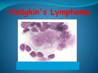
Lymphoma
Lymphoma. Prof DC Stefan Stellenbosch University Cape Town South Africa. LYMPHOMA. Hodgkin’s and non-Hodgkin’s (NHL) 3 rd most common malignancy childhood (incidence 10/mil) NHL 6-7% cancer in children (USA,Europe) NHL most common in sec decade


Lymphoma
E N D
Presentation Transcript
Lymphoma Prof DC Stefan Stellenbosch University Cape Town South Africa
LYMPHOMA • Hodgkin’s and non-Hodgkin’s (NHL) • 3rd most common malignancy childhood (incidence 10/mil) • NHL 6-7% cancer in children (USA,Europe) • NHL most common in sec decade • NHL = most common malignancy in children with HIV (before age 4) • More than 70% NHL will survive at least 5 y with CT (nr of factors)
Non-Hodgkin Lymphoma • All lymphomas not classified as Hodgkin • Originating in cells and organs of immune system • Usually restricted to lymphoid tissue such as lymphnode, spleen ,Peyers patches (could involve BM) • Overlap with ALL pathologically and clinically
NHL • Incidence varies • by age • histological subtype • geographically • Etiology unknown (except Burkitt) • Immunosuppression related to development of NHL, eg. HIV,Wiskott-Aldrich, SCID
Epidemiology NHL • Sex: male: female 2.5:1 • Age-peaks at ages15-19 years • Risks factors: viral (EBV and HIV),radiation, genetic (autoimmune lymph proliferative disorders, severe combined immune deficiency)
Classification • Mature B cell • Burkitt lymphoma 40% • Diffuse large B-cell • Mediastinal large B-cell • Mature T cell • Anaplastic large cell 8-12% • Precursor T lymphoblastic 35% • Precursor B lymphoblastic 2% 15%
WHO Classification • Precursor B-cell neoplasms (leukemia/lymphoma) • Mature(peripheral) B cell neoplasms • Precursor T-cell neoplasms (leukemia/lymphoma) • Mature (peripheral)Tcell neoplasms
CLASSIFICATION NHL • Almost all are high grade-4 groups • 1)lymphoblastic lymphoma • 2)Burkitt’s and Burkitt’s like lymphoma/small non cleaved B-cell lymphoma • 3)diffuse large cell lymphoma • 4)anaplastic large cell lymphoma
Clinical features • Depend on location • BL • Lymphoblastic lymphoma • Anaplastic large cell • Diffuse large cell
Clinical features • Abdomen- 35 % BL ileocecal region • Lymphoma-most common anatomic lesion causing intussusception in children >6 years • Head and neck -13% • Mediastinum 26% svc syndrome • Other sites: peripheral nodes, skin, etc
LYMPHOBLASTIC LYMPHOMA • 30% of NHL • Tumors of thymocytes(T-cell)origin • 75% anterior mediastinal mass • Dyspnea, wheezing,stridor,dysphagia, swelling of the head and neck • +_ involvement lymph node and pleural effusion • Involvement of BM-confusion(25%)
Burkitt’s and Non-Burkitt’s (Small noncleaved cell lymphoma) • 40-50% of NHL • 90% intra-abdominal • Other sites( CNS,peripheral lymphnodes,skin,BM,bone,testis) • B cell origin • 25% contain Epstein Barr virus genomes • TdT enzyme(terminal deoxynucleotidyl transferase)-negative • CALLA (CD10) positive • Characteristic chromosomal translocations
Brief history • Born Enniskillen, Ireland • Education: engineering then medicine • 1938: FRCS, Edinburgh • 1941: Royal Army Medical Corps • 1943: Served with African troops in Kenya • 1946-64: Colonial medical service in Uganda, mainly Mulago hospital • 1957: 2 children seen with swellings in four angles of the jaw … Denis Parsons Burkitt Surgeon & geographical epidemiologist 1911 - 1993
African Burkitt lymphoma • First tumour associated with an oncogenic virus • First tumour successfully treated with combination chemotherapy • Introduced oncology to “tumour lysis syndrome” • Classical chromosomal translocations • Different to Burkitt-type tumours in the west
“Malignant tumours of the jaws in children, primary or secondary, are generally regarded as rare. A sarcoma involving the jaws in African children has recently come to be recognised at Mulago Hospital as a distinctive clinical condition and certainly the commonest malignancy of childhood” “In most cases the tumour started in the region of the alveolar process of a maxilla or the mandible….The tumour grew rapidly grossly distorting the face. Within two or three months of onset symptoms their relatives removed the majority of children from hospital in a moribund condition ” “Deposits other than in the jaws were demonstrated or strongly suspected in 15 patients. In this series the organs most frequently involved were the adrenals, the kidneys and the liver” The British Journal of Surgery, 1958
Burkitt’s observations about occurrence of tumour • In hyperendemic areas, disease found in Black, Asian and European children and less common in children with sickle cell disease • Not at altitudes >3,000 feet 1000 miles south of equator nor at altitudes >5,000 feet at equator • Not when average temperature < 60˚F • Not when annual precipitation < 20 inches • Mostly where falciparum malaria is holoendemic • Does not occur in malarious areas when anti-malarial interventions in place
Burkitt Lymphoma Types: • Immunodeficiency-associated (HIV) • Sporadic • Lymphoid tissues of GIT tract, especially abdomen • Bone marrow (20%) • Endemic • Maxilla, orbit, other facial bones • CNS (30%)
BL – Clinical presentation • Often boys 5-10 years • Extranodal masses, especially lymphoid tissues of GIT tract and upper respiratory tract • Tumour doubling time = ±18hours • High tumour burden • Can cause intussusception, acute abdomen (perforation), ascites, bowel obstruction, airway obstruction,etc. • Spontaneous tumour lysis • Renal failure • Bone marrow failure (if Burkitt ALL) • CNS abnormalities, e.g.. acute flaccid paralysis • Other sites: ovaries, thyroid ,skin, epidural space, bone, pancreas
BURKITT”S • Most nuclei have 1-nucleoli; macrophage at left with debris in cytoplasm
BL - Prognosis • Early stage ~ 95% • Advanced stage ~ 70%
LARGE CELL LYMPHOMA (LCL) • 20-25% of NHL • Anaplastic large cell lymphoma and diffuse large cell lymphoma • Different treatment for diffuse large B cell L and T cell lineage anaplastic large cell L • B-lineage LCL –clinically like small noncleaved L(more localised-mediastinum,BM,CNS) • T-lineage LCL is divided into anaplastic(CD30 positive) and peripheral T cell lymphoma)
Diffuse large B-cell – clinical presentation • Diverse pattern, less predictable • Nodal and extra-nodal • GIT, head and neck, bone, bone marrow, CNS, lymph nodes • Also mediastinal subtype
Anaplastic large cell lymphoma • Nodal and extranodal • Variable presentation Two types: • Primary systemic • Common,fever,weight loss,mediastinum,GIT,bone,peripheral nodes,skin,soft tissue • Easily confused with other systemic diseases • Primary cutaneous • Older patient, often regress spontaneously • >2cm nodules, ulceration,single/multiple
ANAPLASTIC LARGE CELL LYMPHOMA • 90% survival • Short course chemo combined with CNS prophylaxis (only head and neck) • No radiation therapy
ALCL - Prognosis • Systemic: 75-85% • Cutaneous: >90%
DIAGNOSIS • 2 potentially life threatening situations • Superior vena cava sd (mediastinal tumor with airway obstruction)-lymphoblastic lymphoma • Tumor lysis sd (small noncleaved cell NHL)
NHL Staging (St Jude) • I single tumour/nodal mass (not in mediastinum/abdomen) • II single tumour with regional nodes two/more nodes or extranodal masses same side of diaphragm primary GIT tumour • III disease opposite sides of diaphragm all intra-thoracic tumours all extensive abdominal disease paraspinal or epidural tumours • IV + CNS or BM involvement
Burkitt lymphoma • Risk stratification: • Group A, B and C
Life-threatening presentations • Airway compression (careful with sedation) • Pericardial tamponade • Renal failure • Acute abdomen
Staging • Baseline bloods + EBV • HIV • CXR, CT chest • Abdominal sonar, CT abdomen • LP • Bone marrow biopsy • Echo • Histology: FNA, flow cytometry on ascites, biopsy, pleural fluid • Cytogenetics and immunohistochemistry • Bone scan (ALCL,DLBCL)
Treatment • Emergency treatment: • Surgery (abdominal, etc.) • Pericardial tap, pleural tap • Tumour lysis protocol • Steroids/chemo/irradiation • Acute flaccid paralysis • Airway compression
SUDDEN ONSET A 6 year old girl previously healthy presented with severe stridor, several hours before admission and is admitted in ICU being intubated and ventilated.
DIFFERENTIAL DIAGNOSISSUDDEN STRIDOR - croup - bacterial tracheitis - epiglottitis - vocal cord paralysis - foreign body - retropharyngeal abcess - mediastinal cyst - teratoma,etc
INITIAL TREATMENT • NO DIAGNOSIS YET • INHALATIONS AND ANTIBIOTICS- NO RESPONSE
INVESTIGATIONS • CHEST X-RAYS • CT SCAN CHEST • BIOPSY
? LYMPHOMA - EMPIRICAL TREATMENT • STEROIDS • CYCLOPHOSPHAMIDE • VINCRISTINE
COMMENTS • A stridor of abrupt onset is a rather uncommon first manifestation of mediastinal tumors. It is also potentially misleading: such children are easily misdiagnosed with asthma. However, the lack of response to corticosteroids and bronchodilators usually prompts the performing of a chest radiograph which reveals the mediastinal tumor. A CT-scan or MRI provide thereafter further details (1,2,3,4). • Thoracic echography was also proposed as a means of diagnosing anterior mediastinal tumors. It has the merit of easily differentiating between a tumor and a thymic hypertrophy (5). • The life-threatening tracheo - bronchial obstruction in this child made us to start the chemotherapy empirically, in an effort to achieve a rapid reduction in size of the tumor. As a result, the respiratory function improved gradually. In the face of persisting or worsening tracheal obstruction, an alternative approach would be to insert temporary tracheal or bronchial stents (6).