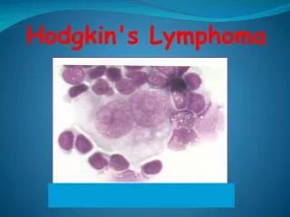
Lymphoma
Lymphoma. Farjah Hassan AlGahtani Assistant Professor, Consultant Hematology Director of Transfusion Medicine and Blood Bank. Overview. Concepts, classification, biology Epidemiology Clinical presentation Diagnosis Staging Three important types of lymphoma. Conceptualizing lymphoma.


Lymphoma
E N D
Presentation Transcript
Lymphoma Farjah Hassan AlGahtani Assistant Professor, Consultant Hematology Director of Transfusion Medicine and Blood Bank
Overview • Concepts, classification, biology • Epidemiology • Clinical presentation • Diagnosis • Staging • Three important types of lymphoma
Conceptualizing lymphoma • neoplasms of lymphoid origin, typically causing lymphadenopathy • leukemia vs lymphoma • lymphomas as clonal expansions of cells at certain developmental stages
ALL CLL Lymphomas MM Neutrophils AML Myeloproliferative disorders Myeloid progenitor Eosinophils Hematopoietic stem cell Basophils Monocytes Platelets Red cells naïve germinal center B-lymphocytes Plasma cells Lymphoid progenitor T-lymphocytes
CLL MM ALL DLBCL, FL, HL B-cell development memory B-cell germinal center B-cell stem cell mature naive B-cell lymphoid progenitor progenitor-B pre-B immature B-cell plasma cell Bone marrow Lymphoid tissue
Clinically useful classification Biologically rational classification • Diseases that have distinct • morphology • immunophenotype • genetic features • clinical features • Diseases that have distinct • clinical features • natural history • prognosis • treatment Classification
Lymphoma classification(2001 WHO) • B-cell neoplasms • precursor • mature • T-cell & NK-cell neoplasms • precursor • mature • Hodgkin lymphoma Non- Hodgkin Lymphomas
Category Survival of untreated patients Curability To treat or not to treat Non-Hodgkin lymphoma Indolent Years Generally not curable Generally defer Rx if asymptomatic Aggressive Months Curable in some Treat Very aggressive Weeks Curable in some Treat Hodgkin lymphoma All types Variable – months to years Curable in most Treat A practical way to think of lymphoma
Mechanisms of lymphomagenesis • Genetic alterations • Infection • Antigen stimulation • Immunosuppression
Epidemiology of lymphomas • 5th most frequently diagnosed cancer in both sexes • males > females • incidence • NHL increasing • Hodgkin lymphoma stable
Incidence of lymphomas in comparison with other cancers in Canada
Risk factors for NHL • immunosuppression or immunodeficiency • connective tissue disease • family history of lymphoma • infectious agents • ionizing radiation
Clinical manifestations • Variable • severity: asymptomatic to extremely ill • time course: evolution over weeks, months, or years • Systemic manifestations • fever, night sweats, weight loss, anorexia, pruritis • Local manifestations • lymphadenopathy, splenomegaly most common • any tissue potentially can be infiltrated
Other complications of lymphoma • bone marrow failure (infiltration) • CNS infiltration • immune hemolysis or thrombocytopenia • compression of structures (eg spinal cord, ureters) • pleural/pericardial effusions, ascites
Diagnosis requires an adequate biopsy • Diagnosis should be biopsy-proven before treatment is initiated • Need enough tissue to assess cells and architecture • open bx vs core needle bx vs FNA
Stage I Stage II Stage III Stage IV Staging of lymphoma A: absence of B symptoms B: fever, night sweats, weight loss
Three common lymphomas • Follicular lymphoma • Diffuse large B-cell lymphoma • Hodgkin lymphoma
Relative frequencies of different lymphomas Non-Hodgkin Lymphomas Diffuse large B-cell Hodgkin lymphoma NHL Follicular Other NHL ~85% of NHL are B-lineage
Follicular lymphoma • most common type of “indolent” lymphoma • usually widespread at presentation • often asymptomatic • not curable (some exceptions) • associated with BCL-2 gene rearrangement [t(14;18)] • cell of origin: germinal center B-cell
defer treatment if asymptomatic (“watch-and-wait”) • several chemotherapy options if symptomatic • median survival: years • despite “indolent” label, morbidity and mortality can be considerable • transformation to aggressive lymphoma can occur
Diffuse large B-cell lymphoma • most common type of “aggressive” lymphoma • usually symptomatic • extranodal involvement is common • cell of origin: germinal center B-cell • treatment should be offered • curable in ~ 40%
Hodgkin lymphoma Thomas Hodgkin (1798-1866)
Epidemiology • ~ 20 000 new cases in North America and Europe every year • Annual incidence 2.7/100 000 per year • Annual mortality only 0.5/100 000 per year • North American lifetime risk – 1/250 to 1/300 • Young adults • 90% in adults 16-65 • Median Age 35 • Slight male predominance • Much less frequent in eastern Asian populations
Associated (etiological?) factors • EBV infection • smaller family size • higher socio-economic status • caucasian > non-caucasian • possible genetic predisposition • other: HIV? occupation? herbicides?
The EBV Association • 3x increased risk Hodgkins with serologically confirmed infectious mononucleosis • EBV genomes detected in ~ 1/3 of Hodgkin lymphoma tissues (developed countries) • Highest proportion mixed cellularity • Population study showed high pre-diagnostic titres of EBV in patients later diagnosed with Hodgkin’s • ?causative – especially in younger patients
Pathology • B cell neoplasm • Unique due to the relative paucity of clonal malignant cells in a background of reactive inflammatory cells • 2 distinct entities • Nodular Lymphocyte predominant HL • L&H cell “popcorn cell” • Classical HL • Reed Sternberg cell • 4 subtypes
Hodgkin lymphoma • cell of origin: germinal centre B-cell • Reed-Sternberg cells (or RS variants) in the affected tissues • most cells in affected lymph node are polyclonal reactive lymphoid cells, not neoplastic cells
Reed Sternberg Cell • “owl’s eye” • 2 nuclear lobes with large inclusion like nucleoli (eosinophilic) • Clear halo around nucleolus (chromatin condensed to nuclear membrane) • Abundant cytoplasm – usually eosinophilic • Lymphocytic and Histiocytic Cell • “popcorn cell” • Polylobated nucleus • Lack of prominent eosinophilic nucleoli • Lack of halo
RS cell and variants classic RS cell lacunar cell popcorn cell (lymphocyte predominance) (mixed cellularity) (nodular sclerosis)
A possible model of pathogenesis loss of apoptosis transforming event(s) EBV? cytokines germinal centre B cell RS cell inflammatory response
Nodular Lymphocyte Predominant Hodgkin’s Lymphoma • 5-10% of patients • “popcorn cell” • Positive for CD 45 • express B-cell associated antigens CD19, CD20, CD22, CD79a, EMA • lack CD15 and CD30 • Background of primarily B lymphocytes +/- histiocytes • Commonly presents early stage (~80%) • 4:1 M:F • slightly higher risk of development of NHL (2% to 5%) • Usually DLBCL • Some treatment differences compared with classical Hodgkin’s
Classical Hodgkin’s Lymphoma • Nodular Sclerosis • Mixed Cellularity • Lymphocyte-depleted • Lymphocyte-rich • CD 15 and CD 30 positive +/- CD 20
Nodular Sclerosis • partially nodular pattern with fibrous bands separating the nodules • lacunar type RS cells - multilobated nuclei and small nucleoli with abundant pale cytoplasm that retracts in formalin-fixed sections producing an empty space • 40%-70% of patients • Commonly present early stage (~70%) • Often confined above the diaphragm • Slight female predominance • Commonly adolescents and young adults
Mixed Cellularity • Classic RS cells common • Background of lymphocytes, eosinophils, plasma cells and histiocytes • 30%-50% of patients • More commonly presents with advanced stage disease, B symptoms • Pediatric and older patients
Lymphocyte-depleted • Classic RS cells with hypocellular fibrotic or reticular background • Presents more commonly in older patients • Commonly advanced stage • Less common involvement of peripheral nodes and mediastinum • Lymphocye-rich • Similar to NLPHL but has classical immunophenotype
Clinical Presentation • Painless lymphadenopathy • Contiguous spread between lymphoid regions • Usually begins supra diaphragmatically • Regional sub diaphragmatic disease < 10% • Symptoms associated with compressive effect • *mediastinal mass • Abdominal/inguinal • “B symptoms” • Wt loss > 10% over 6 months • Persistent fever >38.2 • Drenching night sweats • Puritis
Weird and wonderful… • Alcohol induced pain • Nephrotic syndrome • paraneoplastic secondary to lymphokines • Dermatologic • ichthyosis, acrokeratosis (Bazex syndrome), urticaria, erythema multiforme, erythema nodosum, necrotizing lesions, hyperpigmentation, and skin infiltration
Neurologic • cerebellar degeneration, chorea, neuromyotonia, limbic encephalitis, subacute sensory neuronopathy, subacute lower motor neuronopathy, and the stiff man syndrome • Cholestatic liver disease • Hypercalcemia
Modified Ann Arbour Staging System • I • Single lymph node region (I) • or one extralymphatic site (IE) • II • Two or more lymph node regions, same side of the diaphragm (II) • or local extralymphatic extension plus one or more lymph node regions same side of the diaphragm (IIE)
III • Lymph node regions on both sides of the diaphragm (III) • Which may be accompanied by local extralymphatic extension (IIIE) • IV • Diffuse involvement of one or more extralymphatic organs or sites
A = no B symptoms • B = atleast one of • Unexplained weight loss > 10% during preceding 6 months • Recurrent unexplained fever > 38 • Recurrent night sweats • Bulky disease • Single mass > 10 cm largest diameter
Staging Evaluation • Pathology Review • History looking for B symptoms or other symptoms suggesting systemic disease • Physical for lymphadenopathy and organomegaly • CBC and ESR • Cr, ALP, LDH, bili, Ca, AST, albumin, SPEP • CXR – PA and lat • CT neck, thorax, abdomen and pelvis
Bone marrow aspirate and biopsy if • B symptoms • WBC < 4 • Hgb <120 (women) 130 (men) • Platelets < 125 • ENT examination if • Stage IA or IIA disease with upper cervical lymph node involvement (supra-hyoid)