Download
1 / 75
750 likes | 934 Vues
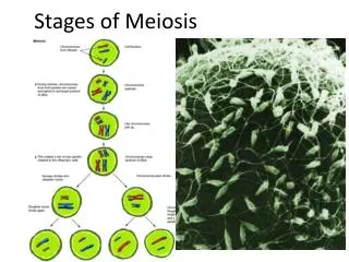
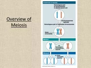
The important bit of meiosis. Cells divide twice First step is essentially mitosis but then they divide again First time there is replication so you end up with a normal number of chromosomes after division – you have 46 pairs which is reduced to 23 pairs

E N D
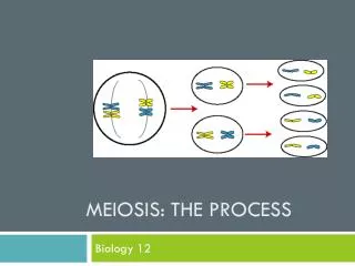
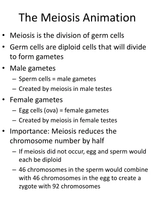
The important bit of meiosis • Cells divide twice • First step is essentially mitosis but then they divide again • First time there is replication so you end up with a normal number of chromosomes after division – you have 46 pairs which is reduced to 23 pairs • From the second division there are now half the number of chromosomes -23 left • Things can go wrong with either step – termed meiosis I and II
Nondisjunction in Meiosis I Paired chromosomes fail to separate. Nondisjunction in Meiosis II Sister chromatids fail to separate.
Monosomy? • Turner’s (XO) • AutosomalTrisomy? • Down’s (Trisomy 21) • Chromosome 21 has a small amount of info on it thus this is compatible with life (also 13,18) • Sex chromosome trisomy • Klinefelters (XXY)
Other genetic abnormalities? • Translocation (Chronic Myeloid Leukemia) • Chromosomes mingle when the meet which can cause errors • Triplet repeats (Huntington’s) • Excess repeats create too much protein e.g. glutamine • Substitution (Sickle cell) • AT subsitution results in abnormal cells • Insertion (Muscular Dystrophy)
What are the 5 pedigrees? • Autosomal Dominant • Autosomal Recessive • X-linked Dominant • X-linked Recessive • Y-linked
Deciphering pedigrees • Look if both sexes equally effected • If no… look if it skips a generation • If it skips a generation, it’s X linked recessive • If not, it’s X linked dominant (affected males don’t have affected sons) • If yes… its autosomal • Look at chances of getting the disease from a diseased parent • ½ = dominant • ¼ = recessive
X-linked dominant • Sex differences • No affected males have affected sons • 1:1 ratio of affected:unaffected daughters
Pedigrees • Autosomal Dominant • BOTH SEXES EQUALLY AFFECTED • Unaffected normal offspring • Affected 1:1 affected:non affected
Autosomal Recessive • BOTH SEXES EQUALLY AFFECTED • Affected individuals usually produce normal (carrier) offspring
X-linked Recessive • Only males effected (pretty much) • Skips a generation
Y-linked • Exclusively affects males • Effected males ALWAYS produce effected males
Complications • (Should probably know two) • Mosaicism • Late onset • Incomplete penetrance • Mitochondrial inheritance
A and a are alleles. p and q are frequencies. A (p) a (q) A (p) AA (p2) Aa (pq) a (q) Aa (pq) aa (q2) Hardy-Weinberg principle Population with an autosomal gene with two alleles (A and a). Frequency of wild type allele A is represented by p. Frequency of defective allele a is represented by q. Since there are only two alleles, p + q = 1. Consider the F1 generation when two heterozygotes (Aa) mate. Chance that offspring is AA is pxp = p2. Chance that offspring is Aa is (pxq) + (pxq) = 2pq. Chance that offspring is aa is qxq = q2.
Applying the Hardy-Weinberg equation p + q = 1 A (p) a (q) A (p) AA (p2) Aa (pq) a (q) Aa (pq) aa (q2) Consider an autosomal recessive that affects 1 in 1600 births. Incidence is q2 = Frequency of allele a is q= Frequency of allele A is p= Carrier (Aa) frequency is 2pq= • 1/1600 • 1/40 • 1-1/40=39/40 • 2x39/40 x 1/40=1/20
Factors required for Hardy-Weinberg equilibrium (know four) • Population is large. • No migration into or out of the population. • Random mating. • Mutation rate remains constant. • No selection of alleles (neither negative not positive).
Transporters • 3 types • Voltage gated • Ligand gated • Mechanically gated (e.g. touch) • Passive, facilitated or active ATPADP
Cl- HCO3- • Be aware of exchangers and anti porters • With vs. anti concentration gradients • May require ATPase – may transfer two ions • Important ones: Cl/HCO3 in RBCs, Ca/Na membrane antiporter, Na/K pump • Or just one: • Ca ATPase extruded out of the cell, Ca ATPase into SR j
Simplified Nernst equation at 37°C 61 mV [ion]out Eion = log [ion]in Z Learn this!! Ratio of Ca outside to in is 10,000:1 EXCLUDES Ca in sarcoplasmic reticulum – it is only the ions next to the membrane that affect the membrane potential
id K+out Ca2+in & K+out 1 2 0 0 Na+in 3 K+out Transmembrane Potential mV -90 4 Na+out K+in Action potential • C • Changing K/Na concentration Ca influx • E.g. Ach nicotinic receptor
Signal types • Paracrine • Endocrine • Autocrine • Direct contact • With three effects: • Change ion balance cascade of effects (e.g.Ca) • Alter gene transcription • Alter existing enzymes via phosphorylation
Second messengers • Learn these ones… • Phospholipase C is activated by G-coupled proteins (Gq alpha units), hydrolyses PIP2 IP3 and DAG • IP3 opens Ca channels on the SR • IP3 is also converted to IP4 to open another Ca channel on the membrane • And this Ca acts to activate further Ca SR release • PLC IP3 IP4 Ca • PIP2 DAG PKC
N C ACTIVE P P OH IP3 OH HO Regulatory domain P N PKC Ca2+ C Catalytic domain INACTIVE IP3-gated Ca2+ channel in intracellular stores DAG, IP3 and Ca2+ and the activation of protein kinase C C=O C=O O O CH2 CH CH2OH DAG
cNMP’S • Adenylatecyclase converts ATPcAMP • Guanylatecyclase converts GTPcGMP • cAMP acts on protein kinase A (amongst others) • cGMP acts on protein kinase G (amongst others) • cGMP and cAMP are common second messengers • The reverse (e.g. cAMPATP) is done by phosphodiesterase • Drugs can impact by inhibiting this process
Receptor types • Intracellular (e.g. steroids) act on the nucleus NOT membrane receptors • Or they may be receptors in the cytoplasm (e.g. NO guanylatecyclase – note NO is a vasodilator) • Ion channels change membrane potential • Na/K • Receptors may have intrinsic function
It’s important to know that this is how ras is activated by growth factors (cancer)
G protein coupled receptors • Ligand binds and stimulates the alpha subunit (all you really need to know) • The alpha subunit has a set function based on its classification • Gs stimulates adenylatecyclase. • Gi inhibits adenylatecyclase. • Gt stimulates cGMPphosphodiesterase. • Gq stimulates phospholipase C.
Acetylcholine – parasymathetic ns • There are five muscarinic acetylcholine receptor subtypes. • M1, M3 and M5 couple through Gq to stimulate phospholipase C. • M2 couples through Gi to open a K+-channel. • M4 couples through Gi to inhibit adenylatecyclase. • PLUS the nicotinic acetylcholine receptor (Na+/K+channel)
Calcium channels • Ca in… • Voltage gated Ca channels (membrane) • IP4 gated Ca channels (membrane) • Ca gated Ca channels (sarcoplasmic reticulum) • IP3 gated Ca channels (sarcoplasmic reticulum) • Ca out… • Plasma membrane ATPase • Sarcoplasmic reticulum ATPase • Ca/Na exchanger
Proteins • Haemaglobin is a quaternary struture • 4 globins and a haem • 2 alpha/2 beta structure • Only two things bind to the haem… • O2 • CO (NOT CO2 – this binds to the globin)
Postiveallosterism • Relaxed haem binds O2 more readily • This is essential for it to be able to unload O2 at tissues
What lowers the affinity of haem for O2? (note this shifts the curve to the RIGHT) • 2,3, BPG • Stabilises deoxygenated Hb • H or CO2 – effect on pH? • Increases acidity – the Boer effect • H binds to Hb and stabilizes dexoygenatedHb • CO2 is converted to HCO3 and binds to Hb (‘carbamation’) - stabalises deoxygenated Hb • CO2+H2O H2CO3 HCO3- + H+
Acid-base balance • Ventilation (breathing) excretes CO2 • Hence hyperventilation respiratory alkalosis • And hypo ventilation respiratory acidosis • You also get metabolic imbalance • Increased acid (e.g. ketones in diabetes) metabolic acidosis • Increase HCO3 metabolic alkalosis • Either way, it comes back to this… • CO2+H2O H2CO3 HCO3- + H+ • Lungs Kidney
Collagen • Triple helix (Left handed) • Glycine: Proline/hydroproline • Or glycine:lysine/hydrolysine • Major part of the extra cellular matrix • Need to know steps of post-translational modification and diseases related to them
Post translational steps • 1: Synthesise alpha chains of pre-pro collagen • (Pre and pro are both precursors) • 2: Hydroxylateproline to hydroxyproline • (requires vitamin C so problem = scurvy) • 3: Hydroxylate lysine residues to hydroxylysine • 4: Glycosylate some hydroxlysine • These steps are done to enable cross linking and glycosylation gives a more open structure
5: Assemble 3 chains to form procollagen • 6: These zipper together into the triple helix
7: Remove the globular ends by procollagen peptidase, so that fibrils can be formed • (lack of this ethlersdanlos (hyper mobile joints, stretchy skin) • 8: Cross link fibrils (lysines and hydroxlysines) to form collagen. • Requires lysyloxidase: defect lathyrism (curved spine, aortic aneurysm, dislocations)
OsteogenesisImperfecta • Glycinebulky amino acid, so type 1 collagen (bone) can’t fold correctly and is unstable • Fractures • NOT a post translational disease (primary structure)
Blood • Haematocrit is just the red blood cell cell content (usually 40-45%)
Haematopoiesis (making blood) • EPO driven (produced by the kidney)
Anemia • Lots of causes • Can be a symptom of bleeding anywhere in the body (ulcers, malignancy) • Symptoms: • Pallor (sign?) • Tiredness • Fainting • Light headedness • Dyspnoea
How does the body respond? • 2,3 BPG • Redistribute blood to important places • Produce reticulocytes (immature RBCs, limited use – note LARGER)
3 Classifications • Normocytic, normochromic • Cells are the same, just less • So blood less – either Acute Blood Loss, or Anemia of Chronic Disease • Microcytic, hypochromic • Little girly cells • No iron • Menstruation • Macrocytic, normochromic • BIG cells (may be reticulocytes) • Vitamin B12/Folate deficiency • Intrinsic factor required for uptake – any question involving terminal ilium, think anemia!
4 stages following vascular injury • 1: Vascular constriction • Near instant contraction • Due to inhibited local production of NO and prostacylcin • Usual, relaxed state comes back to pathways…
2: Platelet plug • Collagen and vWF exposed by damage • Platelets bind and activate • Reinforcing : bindingthromboxane A2 and ADP release (platelet activator/aggregators) • Again, we’ve done the pathways…
PLA is converted to PI – COX1 converts this to thromboxane… which does what? • Aspirin inhibits COX1 blood thinner. Why is this permanent?
3: Clotting • Collagen starts intrinsic clotting • Thromboplastin/tissue damage starts extrinsic (slower) • Both of these activate factor X • Which converts ProThrombinThrombin • Which converts Fibrinogen Fibrin • Forms the clot