Download
1 / 25
250 likes | 370 Vues
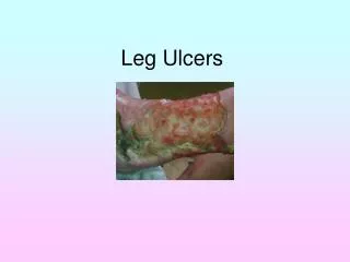
Leg Ulcers. Barry Gibson-Smith Anniesland Medical Practice. Case 1. A 63year old presents with an 8 week history of a non healing area on her shin. She describes that it started after she bumped her leg on a chair. She has been dressing it herself.

E N D
Leg Ulcers Barry Gibson-Smith Anniesland Medical Practice
Case 1 • A 63year old presents with an 8 week history of a non healing area on her shin. She describes that it started after she bumped her leg on a chair. She has been dressing it herself. • What more from her history do you want to know?
History • Ulcer – painful, discharge, smell, itchy, change in size, • PMHx –Venous disease, previous DVT, • Arterial disease – IHD, PVD, CVA • Diabetic • Auto-immune – RA, SLE, Inflam BD, • Cancer – general health • Renal disease • Medication – new? immunosuppressants
Case1 - A 63year old • Established from Hx • Venous veins • Obesity • Poor mobility Your working diagnosis is Venous Ulceration. What signs in the leg would support this?
Venous ulceration Signs may include: • Common site is the medial lower aspect of the calf especially over the malleolus. • Varicose veins • Chronic lymphoedema • Pigmentation • Stasis dermatitis (scratched, dry or weeping patches) • Atrophie blanche (scarring with prominent tortuous capillaries) • Lipodermatosclerosis (firm to hard induration) • Multiple ulcers may occur.
Case1 - A 63year old What are you going to do for this lady who you suspect has venous ulceration of her lower leg?
Case1 - A 63year old • Ankle Brachial Pressure Index (ABPI) by using a Doppler probe to measure pressure in the arm and the ankle. The normal value 0.92 to 1.3. • Peripheral pulses not reliable • ABPI is less than 0.9, there is likely to be arterial disease. Levels of less than 0.5 indicate severe arterial disease. • Caution in diabetics
Case1 - A 63year old • Treatment of/with • Cellulitis • Oedema • Leg elevation • Improve mobility • Dermatitis • Compression bandaging • Aspirin and pentoxyphyline • Avoid – sensitisers, antimicrobials, swabbing, fancy dressings
Case2 - A 58 year old man • Heavy Smoker • Hx of IHD • Hx of intermittent claudication • Ulcer for 5 weeks on toe. • Suspect Arterial Ulcer • What signs would support this?
Arterial Ulceration • Signs- • Usually feet, toes, heels involved and painful • Punched out appearance • Associated with cold white or bluish, shiny feet. • Poor peripheral pulses. • Do ABPI
His ABPI is 0.8 What will you do for him?
Arterial Ulceration • Referral • CVS risk assessment – aspirin, BP, CKD, cholesterol, ?diabetes • Exercise – collateral circulation • Simple dressings • Analgesia • Warning advice - gangrene
Case 3 • A 47 year old lady with 5 year Hx of Rheumatoid arthritis currently on DMARDs therapy. Develops ulcer on lower leg after simple knock. • What type of ulcers present in this group?
Case 3 Ulcers in RA • Pyoderma gangrenosum • Vasculitis • Risk of arterial ulcers • Cellulitis and associated ulceration • Skin cancer ulcers from use of DMARDs
Pyoderma gangrenosum Vasculitis
Case 3 • Importance of prompt referral and secondary care involvement • Likely to require high dose steroids • Admission required if progressing rapidly
Case 4 • An elderly lady presents with a relatively painless ulcer on her lower leg that has been slowly enlarging over several months. What are your differential diagnoses?
Case 4 • Basal cell carcinoma • Squamous cell carcinoma • Amelanotic melanoma • Requires referral for biopsy and excision • Beware of non-healing ulcers
Case 5 A 22year old single mother girl presents with 8 week history of ulcerated area on shin. Lesion is an unusual shape, what diagnosis should be considered and what signs would support it?
Case 5- Dermatitis Artefacta • Bizarre shape depending on methods used to injure the skin • Linear or geometric pattern • Lesions at different stages and range from red patches, swelling, blisters, denuded areas, crusts, cuts, burns, and scars. • Lesions emerge quickly (overnight) without any prior symptoms or signs • Accessible sites such as hands, arms, buttocks, lower legs
Case 6 • A 52 year old diabetic patient presents with a painless ulcer on the sole of his foot. What type ulcers can diabetics suffer from?
Case 6 – Diabetic ulcers • Neuropathic ulcers • Arterial • Venous • Necrobiosis lipoidica • Importance of ABPI and examination • Involvement of diabetic specialist nurses and podiatrist at early stage.
Conclusion • Leg ulcers are a common presentation in practice and require a multi-disciplinary approach • Importance of careful history and examination • Importance of ABPI assessment • Importance of biopsy for non healing ulcerated lesions