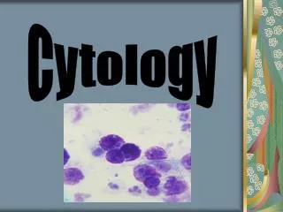
Cytology
Cytology. Kristin M. Canga, RVT References: Bassert and Thomas: Clinical Textbook for Veterinary Technicians , 8 th edition Hendrix and Sirois : Laboratory Procedures for Veterinary Technicians , 5 th edition . Reading Assignments. Laboratory Procedures book: Chapter 9, pp: 287-336


Cytology
E N D
Presentation Transcript
Cytology • Kristin M. Canga, RVT • References: Bassert and Thomas: Clinical Textbook for Veterinary Technicians, 8th edition • Hendrix and Sirois: Laboratory Procedures for Veterinary Technicians, 5th edition
ReadingAssignments • Laboratory Procedures book: Chapter 9, • pp: 287-336 • CTVT: • Pp: 374 – 375, Vaginal Cytology • Pp: 418 – 422, Cytology through end of chapter • Labs for this chapter: • Ear Cytology • Vaginal Cytology • Estrous Cycle cell project
Cytology - Definition • Microscopic examination of individual cells or a small group of cells that have been ________________________ from a tissue or structure. • Cells come from: • _____________________________ • _____________________________ • _____________________________ • _____________________________ Mast Cell Tumor
Histopathology • Greek – • Histos – _____________________________ • Pathos – _____________________________ • Histopathology observes cells in relation to other cells and evaluates the architecture of those tissues. • Involves complex steps and specialized equipment • Use of _____________________________ • Studies the manifestation of _____________________________ • Microscopic _____________________________changes in tissue • More invasive – surgical, biopsy
Why do we do cytology? • Differentiate: • _____________________________ lesion (non-tumor) • _____________________________mass (tumor) • Rapid results • Less expensive • Not as invasive • May be more helpful for certain samples • Mast cell tumor • Not as a replacement for histopathology…adjunct diagnostic tool.
How do we collect samples? • 1: ___________________ • 2: ___________________ • 3: ___________________ • 4: ___________________collection (aspirate and non-aspirate) • 5: ___________________ • 6: ___________________ • 7: ___________________
1: Swabs • Collected when imprints, scrapings and aspirates cannot be made. • Can be taken from ___________________, nasal passages, rectum, ___________________, ___________________ or vagina. • Cells collected are those that have exfoliated from the surface. • You can identify: Yeast, bacteria, WBC, RBC, ear mites, other parasites, fungi,neoplasticcells and epithelial cells.
Swabs – Sample Collection • Materials – Sterile cotton swab, clean glass slide, 0.9% sterile saline, Diff-Quick • Technique: • Pre-moisten a swab with saline (Optional in some cases) • Place pre-moistened swab into/onto the cavity • Use gentle pressure to avoid damage to cells • Gently roll swab onto clean glass slide in a single stroke in 2-3 rows down the length of the slide • Allow to air dry (wave in air) • Apply heat if an ear swab • Stain with Diff-Quik
2: Scrapings • Taken from___________________ lesions on animal • Prepared from tissues collected during___________________ or surgery • Collect many cells from tissue • Advantageous when lesion is firm or yields few cells • Disadvantages – difficult to collect and only ___________________ • Used to reflect a secondary bacterial infection, external parasites, or inflammation-induced tissue dysplasia
Technique • External Parasites: • Supplies: Scalpel blade, mineral oil, slide, gloves • Pinch are of crusted or hairless skin between fingers • Scrape the surface at a 45, or 90 degree angle until capillary oozing (light bleeding) • Place on clean clean slide with mineral oil and examine under microscope • Perform on more than one area if multiple lesions • What external parasites can we find with a skin scraping?
Biopsy Scrapings • Biopsy samples: • Supplies: Scalpel blade, clean slide, stain, gloves • Expose fresh edge of tissue • Blot sample to its nearly dry • Hold blade at 90 degree angle and scrap across the tissue • Spread onto a clean slide – if too thick make a compression slide • Air dry • Stain
3: Imprints (Impression Smear) • Prepared from external lesions on the___________________ animal or from tissues removed during___________________or ___________________ • Collects ___________________ cells than scrapings • Greater amount of___________________ • May hinder the veterinarian in making an accurate diagnosis of___________________ • You may find: Mast cells, bacteria, Yeast, WBC’s, RBC’s, neoplastic cells
Tzanch (Tzanck) Preparation • Imprint preparation – external lesions • Procedure: • 4-6 clean glass slides • Slide 1 – touch to un-prepped lesion • Slide 2 – Prep with saline, gently debride and lightly clean lesion, then touch slide to lesion • Slide 3 – Fully debride lesion and re-imprint with slide • Slide 4 – Imprint the underside of scab if present • Air dry all slides • Stain
Imprint Sample • Taken from tissues collected during surgery or necropsy • Procedure: • Expose a fresh edge on a small piece of tissue • Blood and tissue should be first removed from the surface of the lesion • Touch the tissue repeatedly in rows in single file on a clean slide • Air dry the slide before staining • Consider making multiple slides
4: Fine Needle Collection • Collected from___________________ • Lymph nodes, nodular lesions and internal organs • Advantage: Avoid superficial___________________ • Disadvantage: Fewer cells collected • Methods: • ___________________ • ___________________ • Supplies: • Gloves • Syringe • Needle • Slide • Stain • Surgical prep/alcohol
SitePreparation • Surgical preparation • If sample collected from: • ___________________ • ___________________ • Non-surgical preparation – any other sample • Clean area with alcohol swab
Fine Needle Aspirate (FNA) • Technique: • 21-25 –gauge needle (22g commonly used) • 3-20 ml syringe (12 ml is useful size) • Stabilize mass • Insert needle • Retract plunger to create negative pressure and draw cells • Redirect needle several times • Maintain negative pressure to prevent contamination in lg. masses • Do not exit the mass • Remove needle from mass • Remove syringe from needle • Fill syringe with air • Reattach needle • Gently force sample from needle onto clean slide • Make several slides if possible
Non-aspirate Procedure • ___________________ technique • Stabilize mass • Insert needle (22 gauge) • May have syringe barrel attached without the plunger • Redirect needle several times • Do not exit the mass • https://www.youtube.com/watch?v=0eH5NRzK7QE • Remove needle from mass • Remove syringe barrel (if used) from needle • Fill syringe with air • Re-attach needle • Expel material onto clean glass slide • Repeat
5: Tissue Biopsy • Sampling of a piece of tissue for ___________________and/or___________________ examination • Techniques: Abrasion with blade, needle aspiration, excision and endoscope-guided biopsy. • Varies by___________________ • Consider: location, accessibility and nature • Preparation: • Clip hair • Do NOT cleanse or scrub the site or disrupt scales, crusts or surface debris. • May offer valuable diagnostic information
Tissue Biopsy • Once specimen is collected: • Gently remove specimen by grasping margin of tissue with fine forceps • If collected by endoscopy flush specimen with sterile saline from tip of endoscope • Blot on paper towel to remove excess blood • Place on small piece of wooden tongue depressor and allow to dry • Immerse or float specimen with tongue depressor specimen side down in fixative
Tissue Biopsy - Histopathology • If for histopathologic examination tissues are often fixed in 10% __________________ phosphate-buffered___________________. • Tissue no > than 1 cm • Fluid jar should contain ____xthe specimen’s volume • Large tissues can be transferred to a smaller jar with less formalin after fixed for 24 hours.
Wedge Biopsy • Elliptic specimen commonly obtained with a scalpel • Advantage:___________________ specimen • Excise entire lesion or the wedge is taken from an area of the lesion • Excise through___________________ zone to normal tissue • Pathology technician can trim specimen to provide the pathologist with a slide showing___________________ tissue,___________________ zone and___________________ tissue.
Punch Biopsy • Advantage: speed and procedure • Keyes cutaneous biopsy punches (4, 6, 8-mm) • 6 / 8 mm punches require one or two sutures • 2-3 specimens should be collected • If multiple lesions samples should be collected from the various lesions
Punch Biopsy Procedure • Clip biopsy site being careful to not cause skin inflammation • Select appropriate biopsy punch • Have suture ready if necessary • Gently rotate the biopsy punch in one direction until the punch blade has sectioned the tissue • Grasp the margin of tissue with a pair of fine forceps or flush tissue with saline onto a small piece of wooden tongue depressor • Allow the tissue to dry or blot on paper towel • Place the tissue into a formalin jar, specimen side down on a tongue depressor • Changes in specimen can occur within 1 minute after the biopsy is obtained • Specimens should be allow to fix for at least 24 hours before processing. • Specimens should remain at room temperature for at least 6 hours before exposing to extreme cold.
6: Centesis • Definition: Introduction of a___________________ into any body cavity or organ for the purpose of removing ___________________ • ____________________________________ – collection of fluid from abdominal cavity • ____________________________________ – collection of fluid from the thoracic cavity • ____________________________________ – aspiration of urine from the urinary bladder • ____________________________________ – aspiration of the pericardial sac to retrieve fluid • ____________________________________ – fluid collection from around the spinal column, from within joints and from around the eye
Centesis • Thoracocentesis • Animal in standing position • Needle inserted into the 7th or 8thintercostal space along the cranial aspect of the rib • Abdominocentesis • May be performed in a standing animal or with pt. in lateral recumbency. • Needle introduced into the ventral abdomen to the right of the midline, 1-2 cm caudal to umbilicus
Centesis - Procedures • For peritoneal or pleural fluid the site should be aseptically prepared the site and equipment • A portion of collected fluid should be collected into an EDTA tube to prevent clotting. • 21-gauge needle attached to a 60 ml syringe • Prepare several smears from fluid as soon as it is collected • Record total volume collected, color and turbidity • Determine total nucleated cell counts, cell types, and their morphologic characteristics • General anesthesia required for some procedures such as collection of cerebrospinal fluid
Centesis – Color and Turbidity • Influenced by __________________ concentration and cell numbers • Discoloration may be from contamination with blood, recent or old hemorrhage, inflammation or both. • Perforation of superficial vessels during collection may result in reddish discoloration. • Peripheral blood contamination or recent hemorrhage result in clear supernatant and red (erythrocyte rich) sediment after centrifugation. • Inflammation may cause a degree of turbidity reflecting leukocyte numbers. • Color = off-white, cream, red-cream or dirty brown
7: Transtracheal/ Bronchial Wash • Cytologic evaluation of samples obtained from trachea, bronchi, or bronchioles • May assist with diagnosis of____________________________ disease of animals • ____________________________________ Technique • Involves insertion of needle into trachea through cricothyroid membrane to infuse saline and collect fluid when the animal coughs • Laryngeal area is clipped and aseptically prepared • Local anesthetic – 2% Lidocaine • ____________________________________ Technique • Placing an endotracheal tube in an anesthetized patient and collecting fluid through a jugular or urinary catheter • Preferred in small and/or fractious animals • Animal is lightly anesthetized
Transtracheal Wash - video • http://www.youtube.com/watch?v=QOhGoSgMB5c
Bronchoalveolar lavage / Nasal Flush • Bronchoalveolar lavage – orotracheal technique used to collect samples from the lower respiratory tract. • Bronchoscopy is preferred method but requires bronchoscope • Nasal flush is used for collecting cytologic samples from the nasal cavity • Investigates diseases of the upper airway • Initially, only a small amount of saline infused will be harvested • Cells of interest may be present if the animal coughs after initial collection
Sample Preparation – Orotracheal • All fluid released during coughing should be collected • Place fluid in a sterile tube with notation on site of collection • If sample contains a small amount of mucus centrifuge at a low speed and prepare smears. • If sample contains a large amount of mucus it may not need to be centrifuged. • Nucleated cell counts are not generally performed, but cell numbers are subjectively recorded • Mucus often appears as eosinophilic to purple strands. • Epithelial cells are principal cell type
Smear Preparations – solid masses • Several methods • Experience level of person preparing and characteristics of the sample influence choice of technique • Combination of techniques is suggested: • ____________________________________ Preparation • ____________________________________ Technique • ____________________________________ Smear
Compression “Squash” Preparation • Transfer sample to clean slide near the frosted edge and toward the middle of the slide. • Gently placing a second slide (spreader slide) over aspirate perpendicular to the first slide. • Allow the sample to spread for a few seconds. • Spreader slide (slide 2) is quickly and smoothly slid across prep slide; do not place pressure on spreader of slide. • Air dry spreader slide (slide 2) and stain • Modification: Less tendency to rupture cells • Lay second slide over aspirate, rotate second slide 45 degrees, lift upward.
Combination Technique • Spray sample onto the middle of a clean, glass microscope slide (prep slide) • Place the prep slide on a flat surface and pull another slide (spreader slide) backward at a 45 degree angle to the first slide until it makes contact with approximately 1/3 of the aspirate. • Spread forward as if making a blood smear • The spreader slide is then placed horizontally over the back third of the aspirate at a right angle to the prep slide. • The weight of the slide usually spreads the material • If needed, slide the spreader slide across the prep slide in a quick, smooth motion. • It is important to leave the center portion unspread. This allows for viewing of a high concentration of cells.
Starfish Smear • Transfer sample to the center of a clean slide • Use the tip of a needle to “drag” the sample outward from the center with the point of the needle in a starfish shape • Vary the length and direction of each “drag” through the sample • Does not tend to damage cells • Air dry the slide before staining by gently waving it in the air
Preparation of smears from fluid samples • Smears should be prepared immediately following collection • Collect in ______________ tubes • Smear technique is influenced by cellularity, viscosity, and homogeneity of fluid. • Line smear • Concentrates cells when fluid cannot be concentrated by centrifugation (fluids of low cellularity and translucent) • Place drop of fluid on a clean glass slide • Use blood smear technique except spreading slide is raised directly upward ¾ of the way through the smear = a line of much higher concentrated cells. • Wedge smear – like blood smear
How do we fix and stain our samples? • Advantageous to incorporate a separate cellular fixative • Preferred 95% methanol • Must be fresh and not contaminated with cellular debris • 2-5 minutes (greater is ok) • Romanowsky • Wrights, Giesma, Diff-Quik • Papanicolaou • New Methylene Blue
Romanowsky Stains • Inexpensive and readily available (Diff-quick, Wright’s) • Stain organisms and the cytoplasm of cells • Nucleolar detail sufficient for differentiating neoplasia and inflammation, but Papanicolaou stains are better • Each stain has a unique procedure • Thinner smears, or samples with lower total protein concentration of the fluid generally need less time needed in stain. (And vice versa) • Amount of time in stain (number of dips) will also depend on age of stain. Very variable, be consistent. • Keep stains covered and away from light
Romanowsky Stains Fixative 95% Methanol Eosin – Acidic PH Stains basic components of cells Methylene Blue- Alkaline PH Stains acidic components Distilled Water
New Methylene Blue • Used when critical nuclear detail must be visualized • Stains cytoplasm weakly, if at all • Gives nuclear and nucleolar detail • Useful for when you suspect malignancy
Papanicolaou (Pap stain) • Delicate • Multichromatic stain used for smear preparation • Give excellent nuclear and cytoplasmic detail • Commonly used to diagnose neoplasia • Does not stain cytoplasm as strongly as Romanowsky • Multiple steps and considerable time • 5 dyes in 3 solutions • Specimen must be wet fixed (before cells dry) • Spray with wet fixative or place in ethanol immediately after preparation • With ethanol must be on protein coated slide to prevent cells from falling off slide when immersed. • NOT commonly done in clinic