Download
1 / 9
90 likes | 233 Vues
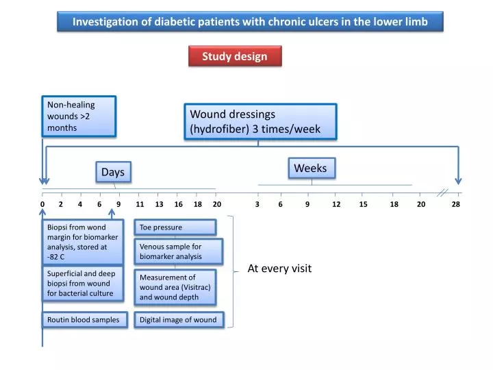
Investigation of diabetic patients with chronic ulcers in the lower limb. Study design. Non-healing wounds >2 months. Wound dressings (hydrofiber) 3 times/week. Weeks. Days. 0. 2. 4. 6. 9. 11. 13. 16. 18. 20. 3. 6. 9. 12. 15. 18. 20. 28.

E N D
Investigation of diabetic patients with chronic ulcers in the lower limb Study design Non-healing wounds >2 months Wound dressings (hydrofiber) 3 times/week Weeks Days 0 2 4 6 9 11 13 16 18 20 3 6 9 12 15 18 20 28 Biopsi from wond margin for biomarker analysis, stored at -82 C Toe pressure Venous sample for biomarker analysis At every visit Superficial and deep biopsi from wound for bacterial culture Measurement of wound area (Visitrac) and wound depth Routin blood samples Digital image of wound
Methods Diabetic patients (type I & II) suffering from a chronic ulcer in the lower limb (failed to heal >2 months after debut) were included in the study. This presentation gives preliminary data from biomarker analyses in 3 patients who failed to heal and 2 patients who healed completely within 15 weeks of observation. Venous blood samples were taken at every visit in pre-chilled tubes (EDTA) and centrifuged in a refrigerated centrifuge, aliquoted to cryotubes and stored at -85oC until analysis. Data are given as mean values. Results are preliminary.
Biochip Multi-spot ECL Technology (MesoscaleDiscovery, USA) This high resolution technique is based on electrochemical stimulation of the electrode surface of multi-spot plates. Antibodies directed against specific biomarkers are attached to a pure non-chargeable carbon surface which eliminates disturbing background activity. Bioluminescence is induced by current and measured by a CCL-camera. Methods
Fibroblast Growth Factor • The FGF family is composed of 23 members. We analysed FGF-2 or basicFGF. Formation of granulation tissue Keratinocytes Chondrocytes Re-epithelialization basicFGF Smooth muscle cells Endothelial cells Tissue remodelling Fibroblasts Mast cells
VEGFR-1 is required for organization of blood vessels. Released upon injury from activated platelets and from macrophages during wound healing Non-healers