Ally, Gina, & Rocky
710 likes | 913 Vues
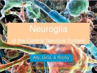
Neuroglia of the Central Nervous System. Ally, Gina, & Rocky. http://www.wordnik.com/words/neuroglia/pronunciations#. Neuroglia. Supportive tissue of the nervous system, including the network of branched cells: Ependymal Astrocytes Oligodendrocytes Microglia. Ependymal Cells.

Ally, Gina, & Rocky
E N D
Presentation Transcript
Neuroglia of the Central Nervous System Ally, Gina, & Rocky http://www.wordnik.com/words/neuroglia/pronunciations#
Neuroglia • Supportive tissue of the nervous system, including the network of branched cells: Ependymal Astrocytes Oligodendrocytes Microglia
Ependymal Cells • Protective layer that lines the brain ventricles and the central canal of the spinal cord • Assists in producing, circulating, and monitoring of cerebrospinal fluid • Epedyma in adults contains stem cells that can divide to produce additional neurons
Astrocytes Largest and most numerous neuroglia in the central nervous system
Functions • Maintains the blood-brain barrier • Isolates the central nervous system from the general circulation • Guides neuron development • Repairs damaged neural tissue • Adjusts the composition of interstitial fluid
Oligodendrocytes • Neuroglia that maintains cellular organization within gray matter and provide a myelin sheath in areas of white matter • Myelin • Insulation around an axon • Consists of multiple layers • Increases impulse rate of the axon
Internodes • Relatively large areas of the axon that is wrapped in myelin Nodes • Small gaps that separate internodes
White matter • Regions of the central nervous system dominated by myelinated axons Gray matter • Region of the central nervous sytem dominated by neuron cell bodies, neuroglia, and unmyelinated axons
Microglia • First form of immune for the central nervous system • 20 percent of the total glial population in the brain • Many fine branches • Capable of migrating through neural tissue
Microglia • Appear in embryonic development • Janitors of the central nervous system • They remain isolated in the neural tissue • Least numerous and smallest Neuroglia in the CNS • Originate from blood cells
Works Cited The free dictionary. (n.d.). Neuroglia. Retrieved February 4, 2010, from http://medical-dictionary.thefreedictionary.com Hybrid medical animation. (n.d.). Glial cells. Retrieved February 4, 2010, from http://www.hybridmedicalanimation.com Martini, F. H. (n.d.). Chapter 12. In Fundamentals of anatomy & physiology (Seventh ed., pp. 384-386). Daryl Fox. (Original work published 2006) The nervous system [French Multipule Sclerosis Research Society]. (n.d.). Retrieved February 4, 2010, from http://images.google.com Neuroglia. (n.d.). Pronunciation. Retrieved February 4, 2010, from http://www.worknik.com
Ion Movements and Electrical Signals By: Caitlin, Drake, Lizzy, Jaylen
Passive Forces Acting Across the Membrane Chemical gradients- Drive sodium ions into the cell. Electrical gradients- Potassium ions leave the cytoplasm more rapidly than sodium ions enter. Current- A movement of charges to eliminate a potential difference. Resistance- A measure of how much the membrane restricts ion movement. Electrochemical gradient- The sum of the chemical and electrical forces acting across the cell membrane for a specific ion.
Active Forces Across the Membrane Passive channels- Membrane channels that are always opened but their permeability varies time to time Active Channels- Membrane channels that open or close when responding to a stimuli, they are activated when opened and inactivated when closed, there are 3 types
Types of Active channels Chemical- open or close to specific chemicals Voltage- open or close according to changes in charges along the membrane Mechanical- Open or close according to physical distortions
Graded Potentials Impulses that cannot travel far from the site of stimulation. “Local Stimulation” Depolarization- any shift from negative resting potential towards 0mV. NA+ ions enter the cell to make it less negative As a result the impulse is sent. Repolarization- process of restoring the cell back to its normal resting potential. The cell becomes more negative.
Graded Potentials Continued Hyperpolarization- cell temporarily becomes more negative than its resting potential. Occurs because too much K+ has leaked out of the cell. Nerve cannot be stimulated until its back to its resting potential.
Action Potentials impulses that travel long distances quickly. All-or-None Principle- once the nerve cell is depolarized past the threshold level the impulse is sent. If it doesn’t reach the threshold, the impulse is not produced. Stimulus either triggers an action potential or it doesn’t. There is no in-between.
Generation of Action Potentials Step 1: depolarization of nerve cell to threshold level. Step 2: NA+ channels open and NA+ ions enter the cell. Creates rapid depolarization. Step 3: NA+ channels close and K+ channels open. K+ ions exit the cell. This starts repolarization. Step 4: Hyperpolarization occurs because too much K+ ions leaked out of the cell. Once the K+ channels are shut, normal resting potential is re-established.
Propagation of Action Potentials propagation- how the impulse travels. Continuous Propagation- occurs on unmyelinated axons, impulse slowly travels down the axon. Saltatory Propagation- on myelinated axons, impulse jumps from node to node. This is much faster than continuous propagation.
Works Cited "Action Potential." Harvard Outreach Animations. Web. 4 Feb. 2010. <http://outreach.mcb.harvard.edu/animations/actionpotential.swf>. Human Physiology. Web. 4 Feb. 2010. <http://people.eku.edu/ritchisong/301notes2.htm>. Matthews, Gary G. "NEUROBIOLOGY." Nuerobiology Animations. Web. 4 Feb. 2010. <http://www.blackwellpublishing.com/matthews/animate.html>. Neuroscience For Kids. Web. 4 Feb. 2010. <http://faculty.washington.edu/chudler/neurok.html>. Society for Neuroscience. Web. 4 Feb. 2010. <http://www.sfn.org/>.
Synaptic Activity Ashleigh Stagg Kevin Williams Kelsey Coulter
Synaptic Activity • Electrical Synapses • Chemical Synapses • Presynaptic- source of action potential • Postsynaptic- Receiving action Potential
Electrical Synapses • Present in some areas of brain • Gap junction- current flows through intercellular channels • Membrane’s potential is changed inhibiting or generating action potential • faster response time
Chemical Synapses • Transmit impulses in 1 direction to a specific location • Fatigue • Synaptic cleft- no intercellular connectivity • Synaptic vesicles • release neurotransmitters
Process of Chemical Synapses • Action Potential arrives and depolarizes synaptic knob • Calcium enters synaptic cleft triggering the release of acetylcholine • Acetylcholine binds to receptors and depolarizes the postsynaptic membrane • Initiates action potential • Acetylcholine is removed through acetyl cholinesterase
Cholinergic Synapses • Cholinergic system includes nerve cells that produce the neurotransmitter acetylcholine • Acetylcholine is a chemical the brain needs to process information and to function normally
Cholinergic cont. • Low acetylcholine levels can lead to Alzheimer’s disease
Neurotransmitters • There are nine chemical compounds belonging to 3 chemical families. • 3 chemical families; Amines, Amino Acids, Peptides. • Chemical released by neurons to stimulate neighboring neurons, allowing impulses to be passed from one cell to the next throughout the nervous system.
Amines • Contains carbon, hydrogen, and nitrogen. • Chemical compounds: • Acetylcholine • Norepinephrine • Dopamine • Seratonin
Amino Acids • Chemical compounds: • Glycine • Glutamic acid • Aspartic acid • Gamma-amino butyric acid
Peptides • Contain at least two amino acids • Chemical Compounds: • Substance P
Works Cited • http://faculty.washington.edu/chudler/genet.html • http://www.elmhurst.edu/~chm/vchembook/662cholinergic2.html • www.thehormoneshop.com/neurotransmitters.htm
Information Processing by Individual Neurons Melanie H. Dezeray H. Oscar R.
Postsynaptic Processing Neurons carry out operations that extract information from sensory receptor. Neurons translate this information into action, imagery and memory. Postsynaptic potentials are graded potentials that develop in the postsynaptic membrane in response to a neurotransmitter. There are two major types.
Excitatory Postsynaptic Potential (EPSP) definition- an electrical change (depolarization) in the membrane of a presynaptic neuron caused by the binding of an excitatory neurotransmitter from a postsynaptic cell to a postsynaptic receptor, making it easier for action potential to generate.
More Information on EPSP there is a temporary depolarization of the postsynaptic membrane they are caused by positively charged ions when the positively charged ions reach the postsynaptic cell, sensitive channels open or close there can be multiple of these which can cause currents and then go into summation EPSP increases the chance of an action potential
During EPSP Na+ (sodium) flows into the synaptic knob causing depolarization (on the left)
Inhibitory Postsynaptic Potential (IPSP) an electrical charge (hyperpolarization) in the membrane of a postsynaptic neuron caused by the binding of an inhibitory neurotransmitter from a presynaptic cell to a postsynaptic receptor; makes it more difficult for a postsynaptic neuron to generate an action potential
More Information on ISPS presynaptic neurons releases neurotransmitters and bind to postsynapic receptors ions channels open and close electrical currents begins and creates a negative postsynaptic membrane postsynaptic cells are inhibited and then it goes into summation IPSP decreases the chance of an action potential
During IPSP K+ (potassium) flows out of the synaptic knob causing hyperpolarization (on the right)
Summation Summation is the ability of skeletal muscle to contract at varying degrees or strength. • There are two types of summation: • Temporal Summation (motor unit) • Spatial Summation (wave)
Temporal Summation Temporal summation is transmission of an impulse by rapid stimulation of one or more pre-synaptic neurons. This stimulation transfers to a motor unit, thus the more motor units stimulated the stronger the contraction.