Chronic Otitis Media
Chronic Otitis Media. Bastaninejad Shahin , MD, ORL & HNS. COM with Cholesteatoma. COM with Cholesteatoma. Epidermal inclusion cyst of the ME or Mastoid cavity Classification: Congenital (usually Ante.Sup.): appear behind or within an intact TM Acquired (usually Post.Sup.):

Chronic Otitis Media
E N D
Presentation Transcript
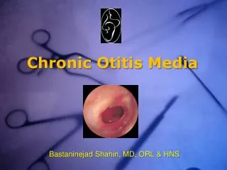
Chronic Otitis Media BastaninejadShahin, MD, ORL & HNS
COM with Cholesteatoma • Epidermal inclusion cyst of the ME or Mastoid cavity • Classification: • Congenital (usually Ante.Sup.): appear behind or within an intact TM • Acquired (usually Post.Sup.): • Otorrhea often malodorous (Mixed>Anaerobes>Aerobes 1 bacteroides, 2 Pseudomonas,...) • Keratin debris in the center of the perforation • Acute flare up may resemble AEO • Vertigo, hearing loss, Facial nerve paralysis,...
Pathogenesis • Congenital: originates from areas of keratinizing epithelium within a small area in the Anterior tympanum • Acquired: • Invagination (ex vacuo theory – in OME or eustachian dysfunction) • Basal cell hyperplasia • Epithelial ingrowth • Squamous metaplasia * *Retraction pocket Cholesteatoma (Sundhoff & Tos)
Complications • Hearing loss through: perforation, ossicular erosion (mainly incus), otic capsule erosions (mainly LSCC, labyrinthine fistula may be found in up to 10% of the cases) • Facial nerve paralysis (acute/chronic) • Tegmen erosion brain hernia or CSF leakage,... • Intracranial infections
Management • Surgical (see next slide for anatomy) • Canal wall down (CWD) • Advantage: recurrence is low • Disadvantage: mastoid cavity problem • Canal wall up (CWU) • Adv.: physiologic position of TM, no mastoid cavity problem • Disadv.: recurrence is high, often nedd second stage exploration
COM without Choles. • Permanent perforation of the TM with or without recurrent infection • Hearing loss regarding to the perforation size: • Small perforation: Low Freq. Air Bone GAP • Large Perforation: Low+High freq. ABG • Bacteriology: usually Mixed>Aerobes>Anaerobes
Management • Medical (mainly topical, in refractory infections use systemic therapy with Ciprofloxacin,...) • Surgery: • Tympanoplasty (dry perforation, less than 25dB ABG) • Tympanomastoidectomy (several episodes of otorrhea, more than 25dB ABG)
Tympanosclerosis • Acellular hyalin and calcified deposits accumulate within the TM and ME submucosa • Pathogenesis: • Consequence of resolved otitis media of trauma • Ossicular fixation may occure (most frequently in the attic head of the malleus and incus)
Management • Management is like COM without cholesteatoma, But ossicular fixation must be corrected simultaneously (except stapedius fixation)