
Introduction & Objectives
hTERT, Survivin and VEGF as antisense targetsin bladder cancer cells: antiproliferative and chemosensitizing effects Meye, S. Fuessel , K. Kraemer, S. Krause, J. Herrmann, S.Ning, Y. Burmeister, M. Kotzsch # , B. Schwenzer, O.W. Hakenberg, M.P. Wirth

Introduction & Objectives
E N D
Presentation Transcript
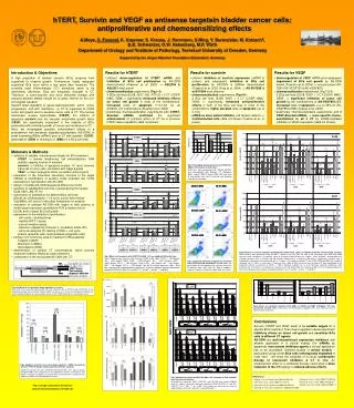
hTERT, Survivin and VEGF as antisense targetsin bladder cancer cells: antiproliferative and chemosensitizing effects • Meye, S. Fuessel, K. Kraemer, S. Krause, J. Herrmann, S.Ning, Y. Burmeister, M. Kotzsch#, • B. Schwenzer, O.W. Hakenberg, M.P. Wirth • Department of Urology and #Institute of Pathology, Technical University of Dresden, Germany • Supported by the Jürgen Manchot Foundation (Düsseldorf, Germany) Introduction & Objectives A high proportion of bladder cancers (BCa) progress from superficial to invasive growth. Furthermore, highly malignant superficial BCa recur within a few years after resection. The currently used chemotherapy (CT) schedules seem to be inefficiently. Moreover, BCa are frequently resistant to CT. Therefore, a tumor-specific and more efficiently therapy with reduced adverse effects should be of great interest for the uro-oncological research. Specific down-regulation of genes associated with tumor onset, progression, and with resistance to CT is supposed to inhibit tumor cell growth and to sensitize tumor cells to CT. The human telomerase reverse transcriptase (hTERT), the inhibitor of apoptosis survivin and the vascular endothelial growth factor (VEGF) are specifically expressed in the majority of BCa, associated with unlimited growth of tumors and resistance to CT. Here, we investigated possible enhancement effects of a pretreatment with antisense oligodeoxynucleotides (AS‑ODN) or small interfering RNAs (siRNA) prior to CT with cisplatin (CDDP), gemcitabine (GEM) or mitomycin C (MMC) in 4 BCa cell lines. • Results for hTERT • efficient down-regulation of hTERT mRNA and inhibition of BCa cell proliferation by AS-ODN demonstrated (Kraemer et al. 2003) ASt2206 & ASt2331 most potent • chemosensitization experiments (Fig.1-3): • 4 BCa cell lines (EJ28, 5637, J82, RT112) + 3 CT (CDDP, MMC, GEM) significantly enhanced inhibitory effects on tumor cell growth in most of the combinations, increased rates of apoptosis (1.3-3.0x) for all ASt2331+CT combinations (Kraemer et al. 2004) • preliminary chemosensitization experiments with hTERT-directed siRNAsconfirmed the significant enhancement of cytotoxic action of CT by a previous hTERT down-regulation (data not shown) • Results for survivin • efficient inhibition of survivin expression (mRNA & protein) and subsequent inhibition of BCa cell proliferation by AS-ODN & siRNAs demonstrated (Fuessel et al. 2003, Ning et al. 2004) AS-SVV286 & si-SVV284 most effective • chemosensitization experiments (Fig.4-6): • 2 BCa cell lines (EJ28, 5637) + 3 CT (CDDP, MMC, GEM) significantly enhanced anti-proliferative effects in both of the BCa cell lines in most of the combinations, highlyelevated rates of apoptosis (up to 3-10x) • siRNA as more potent inhibitor, cell division defects multinucleated cells(data not shown; Fuessel et al., in press) • Results for VEGF • down-regulation of VEGF mRNA and subsequent impairment of BCa cell growth by AS-ODN shown (Foerster et al. 2004) several potent AS-ODN (AS-VEGF723 & AS-VEGF857) • chemosensitization experiments (Fig.7 & 8): • 2 BCa cell lines (EJ28, 5637) + 3 CT (CDDP, MMC, GEM) significant inhibition of tumor cell growth by the combinations of AS-VEGF857+CT, increased rates of apoptosis, e.g. to 35% for AS-VEGF857+MMC (Krause et al. 2004) • preliminary chemosensitization experiments with 3 VEGF-directed siRNAs more specific chemo-sensitizationto all 3 CT by siRNA-mediated inhibition of VEGF expression (data not shown) ** 46% ** 52% *** 55% *** 12% *** 56% *** 38% * 50% * 58% ** 42% * 55% 33% 103% 1 / 2 µg/ml 1 / 2 µg/ml 1 / 2.5ng/ml 1 / 2.5ng/ml 0.33 / 1 µg/ml 0.33 / 1 µg/ml CDDP GEM MMC • Materials & Methods • selection of suitable, overexpressed targets for BCa treatment: • hTERT telomer lengthening, cell immortalization, DNA stability, capping function of telomers • survivin inhibitor of apoptosis protein, 4th most common transcript in tumor cells, correlation with stage & grade • VEGF major angiogenic factor, correlation with progress • calculation of the theoretical secondary structure of the target mRNAs & identification of putative single stranded (ss) motifs accessible for hybridization with AS-ODN • design of multiple AS-ODN targeted at different ss-motifs • selection of suitable BCa cell lines overexpressing the targets: • EJ28, 5637, J82, RT112 • optimization of transfection for different BCa cell lines: • 250nM, 4h, ODN:lipofectin = 1:3 (w/w), serum-free medium • (OptiMEM), diff. points in time after transfection for analysis • evaluation of selected AS-ODN with regard to their potency to inhibit target expression (quantitative PCR & western blot or • ELISA) and to impair BCa cell growth • assessment of the inhibition of proliferation: • cell counts / doubling times • viability (WST-1 assay) • colony formation assay • induction of apoptosis (Annexin V / propidium iodide (PI)) • cell cycle analyses (PI staining of DNA cell cycle • phases, apoptotic cells, multinucleated /polyploidic cells) • testing of CT commonly used for treatment of BCa patients: • Cisplatin (CDDP) • Mitomycin C (MMC) • Gemcitabine (GEM) • determination of suitable CT concentrations, which provoke moderate inhibitory effects as single treatment • combination of the most potent AS-ODN with CT: *** 38% * 69% *** 39% * 38% 77% ** 33% 73% * 53% ** 29% * 61% 85% 58% A) B) Fig.8 Effects of combined treatment with ODNs at 500nM and MMC (0.67µg/ml) 72h after transfection on EJ28 cells.A) Cell counts. B) Percentage of apoptotic cells. Data shown are of one representative experiment. Fig.4 Influence of survivin inhibition combined with 3 different CT agents on viability of the BCa cell lines EJ28 and 5637. Measurements were performed using the WST‑1 viability assay 96h after the start of the transfection with ODN and siRNA constructs and subsequent CT treatment (mean of 5 parallel measurements per sample + mean deviation). Averaged data for isolated treatments with si‑SVV284 and AS‑SVV286 (normalized to si‑luciferase and NS‑K1, respectively) originate from 3 independent experiment series performed with 3 CT agents. The impact of isolated CT with CDDP, GEM and MMC was related to untreated cells (= 100%) in each series. Treatments with siRNA or ODN constructs with or without CT were normalized to the respective controls (si‑luciferase or NS‑K1, = 100%). Residual viabilities (in %) and statistically significant differences after treatment with si‑SVV284+CT in relation to si‑luciferase+CT and AS‑SVV286+CT in relation to NS‑K1+CT are indicated above the bars of the adequate measurements (unpaired Student’s t‑test: (* p0.05, ** p0.01, *** p0.001). 1 / 2 µg/ml 1 / 2 µg/ml 1 / 2.5ng/ml 1 / 2.5ng/ml 0.33 / 1 µg/ml 0.33 / 1 µg/ml Fig.1 Effects of treatment with hTERT-AS-ODN + CT on viability of BCa cell lines. Two CT-doses were used for each cell line. CDDP: EJ28, 5637, RT112 1.0/2.0µg/ml; J82 0.5/1.0µg/ml; MMC: EJ28 0.33/0.67µg/ml, J82 0.67/1.34µg/ml, 5637 0.33/1.0µg/ml, RT112 1.0/1.67µg/ml; GEM: EJ28 1.0/2.5ng/ml, J82 4.0/5.5ng/ml, 5637 2.0/4.0ng/ml, RT112 2.5/4.0ng/ml. The NS-K1 control represents 100%. Error bars represent SD of quadruplicate experiments. Asterisks indicate significant differences between hTERT-AS-ODN+CT and NS+CT (* p0.05, ** p0.01, *** p0.001). CDDP GEM MMC Fig.3Enhanced induction of apotosis by treatment with hTERT-directed AS-ODN+CT in EJ28 cells. The percentages of early apoptotic cells (Annexin V-positive, PI-negative; lower right) and cells died by apoptosis (Annexin V-positive, PI-positive; upper right) are shown. 24h 24h 24h incubation for further CDDP, GEM 24, 48 or 72h 72h 20h Seeding CT Transfection WST WST - - 1 1 assay assay Cell counting Cell counting (4h) 2h 2h 2h Apoptosis detection Apoptosis detection MMC MMC Cell cycle analysis Fig.2 Prolonged doubling times after hTERT-AS-ODN+CT treatment in EJ28 cells. EJ28 cells were pretreated with 250nM ASt2331 and subsequently with different CT (0.33µg/ml MMC, 1µg/ml CDDP, 2.5ng/ml GEM). 96h after start of transfection cells were harvested and doubling times were calculated. Fig.5 Inhibition of cell growth by down-regulation of survivin. EJ28 cells were harvested and counted 72h after treatment with si‑SVV284 and AS‑SVV286 or the respective controls with or without 2.5ng/ml GEM. Cell counts as a measure of cell growth inhibition (A) were related to inhibition of survivin expression (B) at the mRNA level and (C) at the protein level. Conclusions Survivin, hTERT and VEGF seem to be suitable targets for a specific BCa treatment. Their down-regulation causes significant inhibitory effects on tumor cell growth and sensitizes BCa cells to different CT agents. AS-ODN are well-characterized expression inhibitors and already applicable in a clinical setting. For siRNAs as apparently more potent inhibitory agents a clinical application has to be elucidated. Detailed studies in animal models particularly using human BCa cells orthotopically implanted in nude mice will show the suitability of a (local) combination therapy of expression inhibitors & CT in vivo.An enhancement effect of a combined therapy could allow a dose reduction of the CT leading to reduced adverse effects. • References • Forster Y. et al.Cancer Lett. 2004;212:95-103. • Fuessel S. et al. J Urol. 2004;171:2471-6. • Fuessel S. et al.Cancer Lett.;2006;232:243-54. • Kraemer K. et al. Clin Cancer Res.; 2003;9:3794-800. • Kraemer K. et al. J Urol. 2004;172:2023-8. • Krause S. et al. J Urol. 2005;174:328-31. • Ning S. et al. Int J Oncol. 2004;25:1065-71. Fig.6 Apoptosis induction by survivin down-regulation + MMC measured by Annexin V‑FITC/PI double staining and flowcytometric analyses. Treatment of EJ28 cells with si‑SVV284 and AS‑SVV286, resp., with or without 0.67µg/ml MMC were compared to the appropriate controls (si‑luciferase and NS‑K1). Changes in apoptosis (n‑fold increase) of treated cells in comparison to adequate controls are indicated above the bars. Fig.7 Inhibition of viability by VEGF-AS-ODN + CT treatment in EJ28 and 5637 cells 72h after transfection. Untreated cells represent 100%. VEGF-AS- and NS-ODN were used at 500nM. Asterisks indicate significant differences between VEGF-AS-ODN+CT and NS-ODN+CT (** p < 0.01, *** p < 0.001). Data shown are expressed as mean of quadruplicates, error bars represent standard deviations. http://urologie.uniklinikum-dresden.de/ susanne.fuessel@mailbox.tu-dresden.de