The Endocrine System Part B
The Endocrine System Part B. 16. Oxytocin. Oxytocin is a strong stimulant of uterine contraction Regulated by a positive feedback mechanism to oxytocin in the blood This leads to increased intensity of uterine contractions, ending in birth

The Endocrine System Part B
E N D
Presentation Transcript
The Endocrine System Part B 16
Oxytocin • Oxytocin is a strong stimulant of uterine contraction • Regulated by a positive feedback mechanism to oxytocin in the blood • This leads to increased intensity of uterine contractions, ending in birth • Oxytocin triggers milk ejection (“letdown” reflex) in women producing milk
Oxytocin • Synthetic and natural oxytocic drugs are used to induce or hasten labor • Plays a role in sexual arousal and satisfaction in males and nonlactating females
Antidiuretic Hormone (ADH) • ADH helps to avoid dehydration or water overload • Prevents urine formation • Osmoreceptors monitor the solute concentration of the blood • With high solutes, ADH is synthesized and released, thus preserving water • With low solutes, ADH is not released, thus causing water loss from the body • Alcohol inhibits ADH release and causes copious urine output
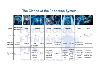
Thyroid Gland • The largest endocrine gland, located in the anterior neck, consists of two lateral lobes connected by a median tissue mass called the isthmus • Composed of follicles that produce the glycoprotein thyroglobulin • Colloid (thyroglobulin + iodine) fills the lumen of the follicles and is the precursor of thyroid hormone • Other endocrine cells, the parafollicular cells, produce the hormone calcitonin
Thyroid Gland Figure 16.7
Thyroid Hormone • Thyroid hormone – the body’s major metabolic hormone • Consists of two closely related iodine-containing compounds • T4 – thyroxine; has two tyrosine molecules plus four bound iodine atoms • T3 – triiodothyronine; has two tyrosines with three bound iodine atoms
Effects of Thyroid Hormone • TH is concerned with: • Glucose oxidation • Increasing metabolic rate • Heat production • TH plays a role in: • Maintaining blood pressure • Regulating tissue growth • Developing skeletal and nervous systems • Maturation and reproductive capabilities
Synthesis of Thyroid Hormone • Thyroglobulin is synthesized and discharged into the lumen • Iodides (I–) are actively taken into the cell, oxidized to iodine (I2), and released into the lumen • Iodine attaches to tyrosine, mediated by peroxidase enzymes, forming T1 (monoiodotyrosine, or MIT), and T2 (diiodotyrosine, or DIT) • Iodinated tyrosines link together to form T3 and T4 • Colloid is then endocytosed and combined with a lysosome, where T3 and T4 are cleaved and diffuse into the bloodstream
Synthesis of Thyroid Hormone Figure 16.8
Transport and Regulation of TH • T4 and T3 bind to thyroxine-binding globulins (TBGs) produced by the liver • Both bind to target receptors, but T3 is ten times more active than T4 • Peripheral tissues convert T4 to T3 • Mechanisms of activity are similar to steroids • Regulation is by negative feedback • Hypothalamic thyrotropin-releasing hormone (TRH) can overcome the negative feedback
Calcitonin • A peptide hormone produced by the parafollicular, or C, cells • Lowers blood calcium levels in children • Antagonist to parathyroid hormone (PTH)
Calcitonin • Calcitonin targets the skeleton, where it: • Inhibits osteoclast activity (and thus bone resorption) and release of calcium from the bone matrix • Stimulates calcium uptake and incorporation into the bone matrix • Regulated by a humoral (calcium ion concentration in the blood) negative feedback mechanism
Parathyroid Glands • Tiny glands embedded in the posterior aspect of the thyroid • Cells are arranged in cords containing oxyphil and chief cells • Chief (principal) cells secrete PTH • PTH (parathormone) regulates calcium balance in the blood
Parathyroid Glands Figure 16.10a
Effects of Parathyroid Hormone • PTH release increases Ca2+ in the blood as it: • Stimulates osteoclasts to digest bone matrix • Enhances the reabsorption of Ca2+ and the secretion of phosphate by the kidneys • Increases absorption of Ca2+ by intestinal mucosal cells • Rising Ca2+ in the blood inhibits PTH release
Effects of Parathyroid Hormone Figure 16.11
Adrenal (Suprarenal) Glands • Adrenal glands – paired, pyramid-shaped organs atop the kidneys • Structurally and functionally, they are two glands in one • Adrenal medulla – nervous tissue that acts as part of the SNS • Adrenal cortex – glandular tissue derived from embryonic mesoderm
Adrenal Cortex • Synthesizes and releases steroid hormones called corticosteroids • Different corticosteroids are produced in each of the three layers • Zona glomerulosa – mineralocorticoids (chiefly aldosterone) • Zona fasciculata – glucocorticoids (chiefly cortisol) • Zona reticularis – gonadocorticoids (chiefly androgens)
Adrenal Cortex Figure 16.12a
Mineralocorticoids • Regulate the electrolyte concentrations of extracellular fluids • Aldosterone – most important mineralocorticoid • Maintains Na+ balance by reducing excretion of sodium from the body • Stimulates reabsorption of Na+ by the kidneys
Mineralocorticoids • Aldosterone secretion is stimulated by: • Rising blood levels of K+ • Low blood Na+ • Decreasing blood volume or pressure
The Four Mechanisms of Aldosterone Secretion • Renin-angiotensin mechanism – kidneys release renin, which is converted into angiotensin II that in turn stimulates aldosterone release • Plasma concentration of sodium and potassium – directly influences the zona glomerulosa cells • ACTH – causes small increases of aldosterone during stress • Atrial natriuretic peptide (ANP) – inhibits activity of the zona glomerulosa
The Four Mechanisms of Aldosterone Secretion Figure 16.13
Glucocorticoids (Cortisol) • Help the body resist stress by: • Keeping blood sugar levels relatively constant • Maintaining blood volume and preventing water shift into tissue • Cortisol provokes: • Gluconeogenesis (formation of glucose from noncarbohydrates) • Rises in blood glucose, fatty acids, and amino acids
Excessive Levels of Glucocorticoids • Excessive levels of glucocorticoids: • Depress cartilage and bone formation • Inhibit inflammation • Depress the immune system • Promote changes in cardiovascular, neural, and gastrointestinal function
Gonadocorticoids (Sex Hormones) • Most gonadocorticoids secreted are androgens (male sex hormones), and the most important one is testosterone • Androgens contribute to: • The onset of puberty • The appearance of secondary sex characteristics • Sex drive in females • Androgens can be converted into estrogens after menopause
Adrenal Medulla • Made up of chromaffin cells that secrete epinephrine and norepinephrine • Secretion of these hormones causes: • Blood glucose levels to rise • Blood vessels to constrict • The heart to beat faster • Blood to be diverted to the brain, heart, and skeletal muscle
Adrenal Medulla • Epinephrine is the more potent stimulator of the heart and metabolic activities • Norepinephrine is more influential on peripheral vasoconstriction and blood pressure
Stress and the Adrenal Gland Figure 16.15
Pancreas • A triangular gland, which has both exocrine and endocrine cells, located behind the stomach • Acinar cells produce an enzyme-rich juice used for digestion (exocrine product) • Pancreatic islets (islets of Langerhans) produce hormones (endocrine products) • The islets contain two major cell types: • Alpha () cells that produce glucagon • Beta () cells that produce insulin
Glucagon • A 29-amino-acid polypeptide hormone that is a potent hyperglycemic agent • Its major target is the liver, where it promotes: • Glycogenolysis – the breakdown of glycogen to glucose • Gluconeogenesis – synthesis of glucose from lactic acid and noncarbohydrates • Release of glucose to the blood from liver cells
Insulin • A 51-amino-acid protein consisting of two amino acid chains linked by disulfide bonds • Synthesized as part of proinsulin and then excised by enzymes, releasing functional insulin • Insulin: • Lowers blood glucose levels • Enhances transport of glucose into body cells • Counters metabolic activity that would enhance blood glucose levels
Effects of Insulin Binding • The insulin receptor is a tyrosine kinase enzyme • After glucose enters a cell, insulin binding triggers enzymatic activity that: • Catalyzes the oxidation of glucose for ATP production • Polymerizes glucose to form glycogen • Converts glucose to fat (particularly in adipose tissue)
Regulation of Blood Glucose Levels • The hyperglycemic effects of glucagon and the hypoglycemic effects of insulin Figure 16.17
Diabetes Mellitus (DM) • Results from hyposecretion or hypoactivity of insulin • The three cardinal signs of DM are: • Polyuria – huge urine output • Polydipsia – excessive thirst • Polyphagia – excessive hunger and food consumption • Hyperinsulinism – excessive insulin secretion, resulting in hypoglycemia
Diabetes Mellitus (DM) Figure 16.18
Gonads: Female • Paired ovaries in the abdominopelvic cavity produce estrogens and progesterone • They are responsible for: • Maturation of the reproductive organs • Appearance of secondary sexual characteristics • Breast development and cyclic changes in the uterine mucosa
Gonads: Male • Testes located in an extra-abdominal sac (scrotum) produce testosterone • Testosterone: • Initiates maturation of male reproductive organs • Causes appearance of secondary sexual characteristics and sex drive • Is necessary for sperm production • Maintains sex organs in their functional state
Pineal Gland • Small gland hanging from the roof of the third ventricle of the brain • Secretory product is melatonin • Melatonin is involved with: • Day/night cycles • Physiological processes that show rhythmic variations (body temperature, sleep, appetite)
Thymus • Lobulated gland located deep to the sternum in the thorax • Major hormonal products are thymopoietins and thymosins • These hormones are essential for the development of the T lymphocytes (T cells) of the immune system
Other Hormone-Producing Structures • Heart – produces atrial natriuretic peptide (ANP), which reduces blood pressure, blood volume, and blood sodium concentration • Gastrointestinal tract – enteroendocrine cells release local-acting digestive hormones • Placenta – releases hormones that influence the course of pregnancy
Other Hormone-Producing Structures • Kidneys – secrete erythropoietin, which signals the production of red blood cells • Skin – produces cholecalciferol, the precursor of vitamin D • Adipose tissue – releases leptin, which is involved in the sensation of satiety, and stimulates increased energy expenditure
Developmental Aspects • Hormone-producing glands arise from all three germ layers • Endocrine glands derived from mesoderm produce steroid hormones • Endocrine organs operate smoothly throughout life • Most endocrine glands show structural changes with age, but hormone production may or may not be affected
Developmental Aspects • Exposure to pesticides, industrial chemicals, arsenic, dioxin, and soil and water pollutants disrupts hormone function • Sex hormones, thyroid hormone, and glucocorticoids are vulnerable to the effects of pollutants • Interference with glucocorticoids may help explain high cancer rates in certain areas
Developmental Aspects • Ovaries undergo significant changes with age and become unresponsive to gonadotropins • Female hormone production declines, the ability to bear children ends, and problems associated with estrogen deficiency (e.g., osteoporosis) begin to occur • Testosterone also diminishes with age, but effect is not usually seen until very old age
Developmental Aspects • GH levels decline with age and this accounts for muscle atrophy with age • Supplemental GH may spur muscle growth, reduce body fat, and help physique • TH declines with age, causing lower basal metabolic rates • PTH levels remain fairly constant with age, and lack of estrogen in women makes them more vulnerable to bone-demineralizing effects of PTH