The Muscular System Part B
300 likes | 574 Vues
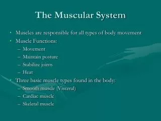
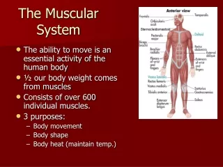
The Muscular System Part B. 10. Muscles of the Anterior Neck and Throat: Suprahyoid. Figure 10.8a. Muscles of the Neck: Head Movements. Major head flexor is the sternocleidomastoid Lateral head movements are accomplished by the sternocleidomastoid and scalene muscles

The Muscular System Part B
E N D
Presentation Transcript
The Muscular System Part B 10
Muscles of the Anterior Neck and Throat: Suprahyoid Figure 10.8a
Muscles of the Neck: Head Movements • Major head flexor is the sternocleidomastoid • Lateral head movements are accomplished by the sternocleidomastoid and scalene muscles • Head extension is accomplished by the deep splenius muscles and aided by the superficial trapezius
Muscles of the Neck: Head Movements Figure 10.9a
Muscles of the Neck: Head Movements Figure 10.9b
Trunk Movements: Deep Back Muscles • The prime mover of back extension is the erector spinae • Erector spinae, or sacrospinalis, muscles consist of three columns on each side of the vertebrae – iliocostalis, longissimus, and spinalis • Lateral bending of the back is accomplished by unilateral contraction of these muscles
Trunk Movements: Deep Back Muscles Figure 10.9d
Muscles of Respiration • The primary function of deep thoracic muscles is to promote movement for breathing • External intercostals – more superficial layer that lifts the rib cage and increases thoracic volume to allow inspiration Figure 10.10a
Muscles of Respiration • Internal intercostals – deeper layer that aids in forced expiration • Diaphragm – most important muscle in inspiration Figure 10.10a
Muscles of Respiration: The Diaphragm Figure 10.10b
Muscles of the Abdominal Wall • The abdominal wall is composed of four paired muscles (internal and external obliques, transversus abdominis, and rectus abdominis), their fasciae, and their aponeuroses • Fascicles of these muscles run at right and oblique angles to one another, giving the abdominal wall added strength
Muscles of the Abdominal Wall • In addition to forming the abdominal wall, these muscles: • Are involved with lateral flexion and rotation of the trunk • Help promote urination, defecation, childbirth, vomiting, coughing, and screaming
Muscles of the Abdominal Wall Figure 10.11a
Muscles of the Abdominal Wall Figure 10.11b
Muscles of the Abdominal Wall Figure 10.11c
Extrinsic Shoulder Muscles • Muscles of the thorax • Anterior: pectoralis major, pectoralis minor, & serratus anterior • Posterior: latissimus dorsi, trapezius muscles, levator scapulae, and rhomboids • These muscles are involved with the movements of the scapula including elevation, depression, rotation, and lateral and medial movements • Prime movers of shoulder elevation are the trapezius and levator scapulae
Extrinsic Shoulder Muscles Figure 10.13a
Extrinsic Shoulder Muscles Figure 10.13b
Muscles Crossing the Shoulder • Prime movers include: • Pectoralis major – arm flexion • Latissimus dorsi and posterior fibers of the deltoid – arm extension • Middle fibers of the deltoid – arm abduction
Muscles Crossing the Shoulder Figure 10.14a
Muscles Crossing the Shoulder Figure 10.14d
Muscles Crossing the Shoulder • Rotator cuff muscles – supraspinatus, infraspinatus, teres minor, and subscapularis • Function mainly to reinforce the capsule of the shoulder • Secondarily act as synergists and fixators
Muscles Crossing the Shoulder Figure 10.14c
Muscles Crossing the Elbow • Forearm extension • The triceps brachii is the prime mover of forearm extension • Forearm flexion • Brachialis and biceps brachii are the chief forearm flexors • The brachioradialis acts as a synergist and helps stabilize the elbow
Muscles of the Forearm • The two functional forearm muscle groups are: those that cause wrist movement, and those that move the fingers and the thumb • These muscles insert via strong ligaments called flexorand extensor retinacula • Most anterior muscles are flexors, and posterior muscles are extensors • The pronator teres and pronator quadratus are not flexors, but pronate the forearm • The supinator muscle is a synergist with the biceps brachii in supinating the forearm
Muscles of the Forearm: Anterior Compartment • These muscles are primarily flexors of the wrist and fingers Figure 10.15a
Muscles of the Forearm: Anterior Compartment Figure 10.15b, c
Muscles of the Forearm: Posterior Compartment • These muscles are primarily extensors of the wrist and fingers Figure 10.16a
Muscles of the Forearm: Posterior Compartment • These muscles are primarily extensors of the wrist and fingers Figure 10.16b