Chapter 6 The Muscular System—Part A
380 likes | 715 Vues
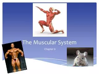
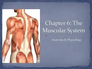
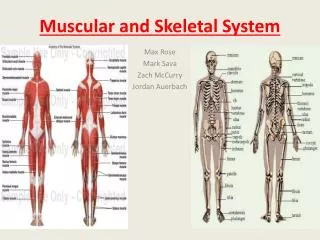
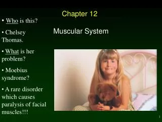
Chapter 6 The Muscular System—Part A. The Muscular System. Muscles are responsible for all types of body movement Three basic muscle types are found in the body Skeletal muscle Cardiac muscle Smooth muscle. Characteristics of Muscles.

Chapter 6 The Muscular System—Part A
E N D
Presentation Transcript
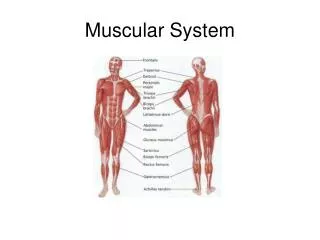
The Muscular System • Muscles are responsible for all types of body movement • Three basic muscle types are found in the body • Skeletal muscle • Cardiac muscle • Smooth muscle
Characteristics of Muscles • Skeletal and smooth muscle cells are elongated(muscle cell = muscle fiber) • Contraction of muscles is due to the movement of microfilaments • All muscles share some terminology • Prefix myo & mysrefers to “muscle” • Prefix sarco refers to “flesh”
Skeletal Muscle Characteristics • Most are attached by tendons to bones • Cells are multinucleate • Striated – have visible banding • Voluntary action– subject to conscious control
Skeletal Muscle & Connective Tissue • Muscle cells are surrounded and bundled by connective tissues • Endomysium – encloses a single muscle fiber • Perimysium– wraps around a fascicle (bundle) of fibers • Epimysium– covers the entire skeletal muscle • Fascia – on the outside of the epimysium Figure 6.1
Skeletal Muscle Attachments • Epimysium blends into a connective tissue attachment • Tendon – cord-like structure • Made of mostly collage fibers • Often cross a joint due to toughness and small size • Aponeuroses– sheet-like structure that attach muscles indirectly to bone, cartilage, or connective tissue coverings • Sites of muscle attachment: • Bones • Cartilages • Connective tissue covering
Smooth Muscle Characteristics • Lacks striations • Spindle-shapedcells • Single nucleus • Involuntary action –no conscious control • Found mainly in the walls of hollow organs Figure 6.2a
Cardiac Muscle Characteristics • Striations present • Usually has a single nucleus • Branching cells • Joined to another muscle cell at an intercalated disc • Involuntary action– no conscious control • Found only in the heart!! Figure 6.2b
Skeletal Muscle Functions • Produce movement • Maintain posture • Stabilize joints • Generate heat
Microscopic Anatomy of Skeletal Muscle • Sarcolemma – specialized plasma membrane • Myofibrils – long organelles inside muscle cell • Sarcoplasmic reticulum – specialized smooth endoplasmic reticulum Figure 6.3a
Microscopic Anatomy of Skeletal Muscle • Myofibrils are aligned to give distinct bands • “I” band = light band • Contains only thin filaments • “A” band = dark band • Contains the entire length of the thick filaments Figure 6.3b
Microscopic Anatomy of Skeletal Muscle • *Note that Myosin and Actin Filaments overlap somewhat
Microscopic Anatomy of Skeletal Muscle • Sarcomere - Contractile unit of a muscle fiber • Organization of the sarcomere • Myofilaments • Thick filaments = myosin filaments • Thin filaments = actin filaments Figure 6.3b
Microscopic Anatomy of Skeletal Muscle • Myosin filaments (Thick) • Composed of the protein myosin • Has ATPase enzymes • Myosin filaments have heads (extensions, or cross bridges) • Actin filaments (Thin) • Composed of the protein actin • Anchored to the Z disc Figure 6.3c
Microscopic Anatomy of Skeletal Muscle • At rest, there is a bare zone that lacksactin filaments called the “H” zone • Sarcoplasmic reticulum (SR) • Stores and releases calcium • Surrounds the myofibrils Figure 6.3d
Stimulation and Contraction of Single Skeletal Muscle Cells • Excitability (also called responsiveness or irritability) – ability to receive and respond to a stimulus • Contractility – ability to shorten when an adequate stimulus is received
Stimulation and Contraction of Single Skeletal Muscle Cells • Extensibility—ability of muscle cells to be stretched • Elasticity—ability to recoil and resume resting length after stretching
The Nerve Stimulus and Action Potential • Skeletal muscles must be stimulatedby a motor neuron (nerve cell) to contract • Motor Unit—one motor neuron and all the skeletal muscle cells stimulated by that neuron Figure 6.4a
The Nerve Stimulus and Action Potential • Neuromuscular junctions • Association site of axon terminal of the motor neuron and muscle Figure 6.5b
Nerve Stimulus to Muscles • Synaptic cleft • Gap between nerve and muscle • Nerve and muscle do not make contact • Area between nerve and muscle is filled with interstitial fluid Figure 6.5b
Transmission of Nerve Impulse to Muscle • Neurotransmitter – chemical released by nerve upon arrival of nerve impulse • The neurotransmitter for skeletal muscle is acetylcholine (ACh) • Acetylcholine attaches to receptors on the sarcolemma • Sarcolemma becomes permeable to sodium(Na+). • Sodium rushes into the cell generating an actionpotential • Once started, muscle contraction cannot be stopped
The Sliding Filament Theory of Muscle Contraction • Activation by nerve causes myosin heads (crossbridges) to attachto binding sites on the thin filament • Myosin heads then bind to the next site of the thin filament and pull them toward the center of the sarcomere • This continued action causes a sliding of the myosin along the actin • The result is that the muscle is shortened (contracted) • Energized by ATP; Requires calcium Figure 6.7