Download
1 / 30
310 likes | 509 Vues
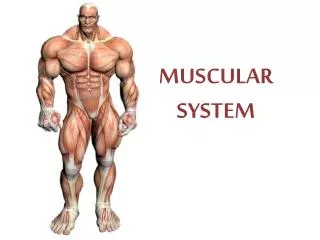
MUSCULAR SYSTEM. TOPIC #4: Muscular System. Muscle Vocabulary Introduction to Muscles Worksheet Microscopic Anatomy of Muscles Vocabulary Quiz Muscle Hodge Podge Skeletal Muscles (36 presentations) Muscle Labels Movement Lab Energy Used for Muscle Contractions Muscular System Diseases

E N D
TOPIC #4: Muscular System • Muscle Vocabulary • Introduction to Muscles Worksheet • Microscopic Anatomy of Muscles • Vocabulary Quiz • Muscle Hodge Podge • Skeletal Muscles (36 presentations) • Muscle Labels • Movement Lab • Energy Used for Muscle Contractions • Muscular System Diseases • Muscular System Review and Crossword • Muscles and Aging
Skeletal muscle presentations • Name • Bone of Origin • Bone of insertion • Joint crosses • Why named? • Action • Antagonistic muscle (if any)
Muscle vocabulary • Threshold stimulus • Motor neuron • Fascicle • Myosin/Actin • Sarcomere • Cholinterase • Acetylcholine • Muscle Cramp • All-or-none Response • Atrophy vs. Atony • Muscle tone • Myostatin • Agonists • Antagonists • Synergists • Fixators
BASIC TRAINING • Latin word mus? Little mouse • 3 types of muscles? Skeletal, cardiac, smooth • Striated? • Tendons? • Housekeeping muscles? • Functions of muscles? • Key words: • Skeletal: skeletal, striated, voluntary • Smooth: involuntary, visceral, non-striated • Cardiac: cardiac, involuntary, striated • Cardiac muscle: HEART • Contracting: muscle shortens
MICROSCOPIC ANATOMY Contains cells, nerve fibers, connective tissues, blood vessels Covered externally by epimysium Bundle of cells, separated by connective tissue sheath Surrounded by perimysium Elongated, multiple cells, multinucleate, has striated appearance Surrounded by endomysium
MUSCLE FIBERS ARE NOT SINGLE CELLS! • They are a FUSION of many cells = myoblasts • Each Muscle Fiber Contains: • An array of myofibrils • Mitochondria – responsible for Cellular Respiration • Convert chemical energy (food) into cellular energy (ATP) • Also called sarcosomes • Many nuclei (multi-nucleate) • Sarcolemma = cell membrane • Sarcoplasmic reticulum = ER • Sarcoplasm?? • Sarcomere…
SARCOMERES • Muscle fibers are STRIATED • Myofibrils are made up parallel filaments (thick & thin) • Striations are created by dark A bands and light I bands • A bands are bisected by the M line • I bands are bisected by the Z disk • Thick filaments produce the dark A band & contain myosin • Thin filaments produce the light I band & contain actin
Entire array of thick and thin filaments between Z disks video
Read pages 166 and 167 WRITE OUT QUESTIONS AND ANSWERS PLEASE! • What split ATP to generate power for muscle contraction? • What links the thick and thin filaments together during muscle contraction? • Why does the light, H zone seem to disappear during muscle contraction? • What is the major role of the SR? • What provides the “go” signal for muscle contraction? Also answer #1 on page 198 and #3 on page 199
#5 Muscle Hodge Podge
Golden Rules of Muscle Activity • All muscles cross at least one joint. • Typically, the bulk of the muscle lies PROXIMAL to the joint crossed. • All muscles have at least 2 attachments: the origin and the insertion. • Muscles can only pull, they never push. • During contraction, the muscle insertion moves toward the origin.
Points of muscle attachment in bone • Origin = less movable bone • Insertion= movable bone; moves toward origin when contracting
Body movement occurs when muscles contract across joints • Flexion, Extension, Hyperextension • Rotation (lateral = away vs. medial = towards) • Abduction (away from midline), Adduction (toward midline) • Circumduction • FOOT: Dorsiflexion, Plantar Flexion, Inversion, Eversion, • Supination, Pronation • Opposition • Elevation, Depression, Retraction, Protraction
Elevation: scapula is raised, “shrugging the shoulders” • Depression: scapula pulls down to a more inferior position • Protraction: scapula moves forward, “hunching the shoulders” • Retraction: scapula moves backward, “squaring the shoulders”
Muscles CANNOT push! • Muscles must work in REVERSE pairs to move the bones. • Immense variety of movement possible • Prime mover: muscle with majority of responsibility of causing movement • Antagonists: oppose/reverse a movement • Synergists: help prime movers by producing same movement or lessening other movements • Fixators: specialized synergists, used for stabilizing purposes
NaaaammmmeThat Muscle! • Muscles are named, based on various characteristics. • Location example: tibialis anterior is located near the front of the tibia bone. • Size: example: maximus means larger & minimus means smaller. (pectoralis major & minor muscles of the chest)
Number of origins/insertions: based on the number of origins, example: quadriceps femoris (which has 4 = quad) • Fiber direction: example: rectus (straight) runs parallel to midline of body. Oblique run slanted to midline of the body. • Muscle shape: example: deltoid means triangular • Muscle action: example: flexor, adductor, extensor
Important Skeletal Muscles • Facial Muscles • Neck Muscles • Anterior Trunk Muscles • Posterior Trunk Muscles • Upper Limb Muscles • Lower Limb Muscles
Muscles of the Human BodyMuscles of the head and neck: The muscles of the face and head can be divided into 3 main categories: (A) muscles of expression – orbicularis, buccinator, frontalis, occipitalis (B) muscles of chewing – masseter, temporalis and (C) muscles of the neck – sternocleidomastoid. Muscles of the Neck and Shoulder:Sternocleidomastoid, Trapezius, Deltoid, Rotator Cuff: is a group of 4 muscles which hold the head of the arm bone in the shoulder joint and attach the arm to the chest. Muscles of the Chest and Back: muscle of the chest includes - Pectoralis Major, Pectoralis Minor and Intercostal Muscles. The muscles of the back include – Trapezius, LatissimusDorsi and Serratus Posterior. Muscles of the Upper Extremity: the muscles of the upper arm include – Coracobrachialis, Biceps, Brachialis and the Triceps muscle. The muscles of the forearm include - PronatorTeres, Extensor DigitorumCommunis and the Flexor Carpi Radialis. Muscles of the hand include - Palmaris Brevis, Abductor DigitiQuinti, Abductor PollicisBrevis and the Flexor PollicisBrevis muscle. Muscles of the Lower Extremity: the muscles of the thigh and shin region include - Quadriceps Muscles, Hamstring Muscles, Tibialis Anterior, Calf Muscles and yhe Extensor DigitorumLongus muscle. The muscles of the foot include - Plantar Aponeurosis, Abductor Hallucis, Flexor DigitorumBrevis and the Abductor DigitiQuinti.
As You Age, Your Muscles… • decrease in size (diameter) • lose myofibrils(especially fast twitch) • contain less ATP & glycogen = less power produced = tend to fatigue more easily • develop Fibrosis (increase in connective tissue) • makes muscles less flexible &inhibits movement • have less blood flowing to them (reduced circulation) • do not heal/repair themselves as well • OVERALL EFFECT: reduced size, strength, endurance
Muscular System Diseases • Usually extremely painful, result in physical impairments (such as an inability to walk). • Nerve damage • Muscular Dystrophy • Myasthenia Gravis • ALS • Cerebral palsy • Torticollis
Page 198 Multiple Choice • 1. c. Light I bands – contain only actin – during contraction, sarcomeres shorten – cause actin filaments to slide together leaving only dark A bands visible. • 3. c. Resistance • 4. b. abducting (maybe a. extending) • 5. all of them! • 6. all but c (vastus do not attach to hip) • 7. all but c (vastus do not move hip) • 8. a. biceps, b. triceps, d. lat
At the Clinic • #1: deltoid, gluteus medius (for big amounts), 2 quad muscles (lat. vastus & rectus femoris)*** • #2: tore gastrocnemius from its insertion point in Achilles tendon • #3: pects major, lats, deltoid, triceps, biceps, traps • #4: Eric utilized glycolysis and anaerobic respiration because lack of oxygen. This makes less ATP which sends the cells into lactic acid fermentation. Lactic acid builds up causing the pain. • #6: lateral flexion of spine causes scoliosis
STUDY! • Functions of muscles • Compare skeletal, smooth and caridac • Microscopic Anatomy of muscles • Attachments to bones • Body movements • Agonist vs. antagonist vs. fixators vs. synergists • Presentations (mostly actions) • Aging • Lab terminology (chewing muscles, etc.) • Diseases


