Chapter 6 The Muscular System—Part C
480 likes | 851 Vues
Chapter 6 The Muscular System—Part C. Muscles and Body Movements. Movement is attained due to a muscle moving an attached bone Muscles are attached to at least two points Origin Attachment to an immoveable bone Insertion Attachment to a movable bone

Chapter 6 The Muscular System—Part C
E N D
Presentation Transcript
Muscles and Body Movements • Movement is attained due to a muscle moving an attached bone • Muscles are attached to at least two points • Origin • Attachment to an immoveable bone • Insertion • Attachment to a movable bone • During contraction, insertion moves toward origin
Types of Ordinary Body Movements • Flexion • Decreases the angle of the joint • Brings 2 bones closer • Ex: hinge joints like knee & elbow • Ex: ball & socket joints like hip & shoulder • Extension • Opposite of flexion • Increases the angle between two bones • Ex: Straightening knee or elbow
Types of Ordinary Body Movements • Hyperextension • Line of appendage is over 180° when straightened • Ex: Tip head posteriorly
Types of Ordinary Body Movements • Rotation • Movement of bone around its longitudinal axis • Common in ball & socket joints • Ex: Move atlas around the axis (shake your head “no”)
Types of Ordinary Body Movements • Abduction • Movement of a limb away from the midline • Adduction • Opposite of abduction • Movement of a limb toward body midline • Circumduction • Combination of flexion, extension, abduction, adduction = circular • Common in Ball & socket joints
Special Movements • Dorsiflexion – • Lifting the foot so that the superior surface approaches the shin • Plantar flexion • Depressing the foot (pointing the toes)
Special Movements • Inversion • Turn sole of foot medially • Eversion • Turn sole of foot laterally
Special Movements • Supination • Forearm rotates laterally so palm faces anteriorly • Radius & Ulna are parallel • Pronation • Forearm rotates medially so palm faces posteriorly • Radius & Ulna form “X” shape
Special Movements • Opposition • Move thumb to touch the tips of other fingers on the same hand
Types of Muscles • Prime Mover – muscle with the major responsibility for a certain movement • Antagonist – muscle that opposes or reverses a prime mover • Muscle can be both: • Biceps – prime mover flexes elbow • Triceps – prime mover extends elbow • Synergist – muscle that aids a prime mover in a movement and helps prevent rotation • Fixator – stabilizes the origin of a prime mover • Allows all tension to move insertion bone
Naming of Skeletal Muscles • Based on direction of muscle fibers • Example: Rectus (straight) • Based on relative size of the muscle • Example: Maximus (largest) Minimus – smallest Longus - long • Based on location of the muscle • Example: many muscles are named for bones Temporalis(temporal bone)
Naming of Skeletal Muscles • Based onnumber of origins • Example: Triceps (three heads) • Bi – 2 • Quad -4 • Based on location of the muscle’s origin and insertion • Example: sterno (on the sternum)
Naming of Skeletal Muscles • Based on the shape of the muscle • Example: deltoid --triangular • Based on the action of the muscle • Examples: flexor and extensor --flexes or extends a bone
Arrangement of Fascicles Figure 6.14
Fascicles • Circular – rings of muscle that open and close areas • Sphincters • Orbicularisoris – around mouth • Convergent – muscles come together to a single insertion point—usually a tendon • Fan or triangle shaped • Pectoralis major • Parallel – length runs parallel to long axis of muscle • Straplike
Fusiform – spindle shaped, long • Biceps brachii • Pennate – fascicles attach obliquely (at a slanted position) to central tendon
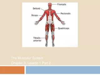
Head and Neck Muscles • Facial muscles • Frontalis—raises eyebrows • Orbicularisoculi—closes eyes, squints, blinks, winks • Orbicularisoris—closes mouth and protrudes the lips • Buccinator—flattens the cheek, chews • Zygomaticus—raises corners of the mouth • Chewing muscles • Masseter—closes the jaw and elevates mandible • Temporalis—synergist of the masseter, closes jaw
Head and Neck Muscles • Neck muscles • Platysma—pulls the corners of the mouth inferiorly • Sternocleidomastoid—flexes the neck, rotates the head
Head and Neck Muscles Figure 6.15
Muscles of Trunk, Shoulder & Arm • Anterior muscles • Pectoralis major—adducts and flexes the humerus • Intercostal muscles • External intercostals—raise rib cage during inhalation • Internal intercostals—depress the rib cage to move air out of the lungs when you exhale forcibly
Anterior Muscles of Trunk, Shoulder, Arm Figure 6.16
Muscles of Trunk, Shoulder, Arm • Muscles of the Abdominal Girdle: • Rectus abdominis—flexes vertebral column and compresses abdominal contents (defecation, childbirth, forced breathing) • External and internal obliques—flex vertebral column; rotate trunk and bend it laterally • Transversusabdominis—compresses abdominal contents
Anterior Muscles of Trunk, Shoulder, Arm Figure 6.17
Muscles of Trunk, Shoulder, Arm • Posterior muscles: • Trapezius—elevates, depresses, adducts, and stabilizes the scapula • Latissimusdorsi—extends and adducts the humerus • Erector spinae—back extension • Quadratuslumborum—flexes the spine laterally • Deltoid—arm abduction
Muscles of the Upper Limb • Biceps brachii—supinates forearm, flexes elbow • Brachialis—elbow flexion; lifts ulna • Brachioradialis—weak muscle • Triceps brachii—elbow extension (antagonist to biceps brachii)
Muscles of the Lower Limb • Gluteus maximus—hip extension • Gluteus medius—hip abduction, steadies pelvis when walking • Iliopsoas—hip flexion, keeps the upper body from falling backward when standing erect • Adductor muscles—adduct the thighs
Muscles of the Pelvis, Hip, Thigh Posterior view of hip and thigh
Muscles of the Lower Limb • Muscles causing movement at the knee joint: • Hamstring group—thigh extension and knee flexion • Biceps femoris • Semimembranosus • Semitendinosus
Muscles of the Lower Limb • Muscles causing movement at the knee joint • Sartorius—flexes the thigh • Quadriceps group—extends the knee • Rectus femoris • Vastus muscles (three)
Muscles of the Pelvis, Hip, Thigh Anterior view of pelvic & thigh muscles
Muscles of the Lower Limb • Muscles causing movement at ankle and foot: • Tibialis anterior—dorsiflexion and foot inversion • Extensor digitorumlongus—toe extension and dorsiflexion of the foot • Fibularis muscles—plantar flexion, everts the foot • Soleus—plantar flexion
Developmental Aspects • Nervous system must mature before muscle control can be achieved. • Gross movements develop before precise control is possible. • Maximum development of nervous system achieved in mid-teens—athletes often notice an increased level of performance.
Homeostatic Imbalances • Muscular Dystrophy – group of inherited muscle-destroying diseases that affect specific muscle groups • Muscles enlarge due to fat and connective tissue deposits, but muscle fibers degenerate • Caused by lack of the protein dystrophin that is found in the sarcolemma • Duchenne’s Muscular Dystrophy • Primarily affects males • Do not live beyond young adult
Homeostatic Imbalances • Myasthenia gravis • Characterized by drooping of eyelids, difficulty in swallowing and talking, generalized muscle weakness • Shortage of acetylcholine receptors at the neuromuscular junction • Autoimmune disease
Homeostatic Imbalances • Effects of Aging • Connective tissue increases, muscle mass decreases • Body weight decreases • Strength decreases • These effects can be countered by regular exercise, including weight training