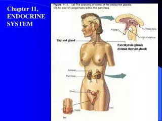
Chapter 6: Muscular System
Chapter 6: Muscular System. Anatomy & Physiology Kasprowicz. The essential function of muscle is contraction – making it responsible for almost all body movement. Types of Muscle Skeletal Cardiac Smooth . Types of Muscle . muscle cells are elongated =

Chapter 6: Muscular System
E N D
Presentation Transcript
Chapter 6: Muscular System Anatomy & Physiology Kasprowicz
The essential function of muscle is contraction – making it responsible for almost all body movement.
Types of Muscle Skeletal Cardiac Smooth Types of Muscle
muscle cells are elongated = • muscle fibers (smooth, skeletal) • contain myofilaments (ability • to contract) • terminology: • myo-, mys- = muscle • sarco- = flesh General Muscle Characteristics
Very large, multinucleated cells • Striated (visible stripes or banding pattern) • Voluntary (conscious) control; can be reflexive too • muscle fibers (cells) are bundled together by strong connective tissues exert great force, but tire easily Skeletal Muscle
Deep fascia Connective Tissues in Skeletal Muscle
Endomysium – around single muscle fiber • Perimysium – around a fascicle (bundle) of fibers • Epimysium – covers the entire skeletal muscle • Deep Fascia – on the outside of the epimysium Connective Tissues in Skeletal Muscles
tendon – dense connective tissue attaching muscle to bone (cord-like) • aponeuroses – attach muscles indirectly to bone,cartilages or connective tissue coverings (sheet-like) • Epimysium blends into a connective tissue attachment Connective Tissues in Skeletal Muscles
Aponeurosis of the external oblique Connective Tissues in Skeletal Muscles
Refer to pg. 272 in your textbook Connective Tissues in Skeletal Muscles
Spindle-shaped cells with one nucleus • no striations • Involuntary (no conscious control) • Found in hollow visceralorgans • Have small amount of endomysium Smooth Muscle
slow, sustained, tireless movement • often layers in opposite directions Smooth Muscle
Branching cells with one nucleus connected by intercalated discs 2) Striated 3) Involuntary (no conscious control) • small amounts of endomysium Cardiac Muscle
Cardiac fibers are arranged in spiral or 8-shaped bundles Cardiac Muscle
Producing movement • Maintaining posture • Stabilizing joints • Support soft tissues • Generating heat • Guard entrances and exits Muscle Functions
Cells are multinucleate • Nuclei are just beneath the sarcolemma (1) sarcolemma - specialized plasma membrane of muscle cells Microscopic Anatomy of Muscle
(2) Cytoplasm filled with myofibrils myofibrils – perfectly aligned, ribbon- like organelles; give muscle fiber its striped appearance Microscopic Anatomy of Muscle
(2) Cytoplasm filled with myofibrils A closer look at the myofibril Light (I) bands Dark (A) bands Microscopic Anatomy of Muscle
(2) Cytoplasm filled with myofibrils Banding Pattern: Light (I) bands contain the Z disc Dark (A) bands contain the H zone M line - holds adjacent filaments together; center of H zone Microscopic Anatomy of Muscle
An even closer look at the myofibril… • Sarcomere - contractile unit of the myofibril - Z disc to Z disc Microscopic Anatomy of Muscle
An even closer look at the myofibril… • Sarcomere Microscopic Anatomy of Muscle
Zooming in on the sarcomere… • Myofilaments- special proteins that cause muscle to contract Two Types: a) myosin (thick filament) b) actin (thin filament) Microscopic Anatomy of Muscle
Two Types: a) myosin (thick filament) • protein with heads that form cross bridges b/t thick & thin filaments during muscle contraction • Contain ATPase enzymes Microscopic Anatomy of Muscle
Two Types: b) actin (thin filament) • Made of the contractile protein actin & some other regulatory proteins Microscopic Anatomy of Muscle
Zooming in around the H zone…. Microscopic Anatomy of Muscle
(5) Sarcoplasmic reticulum (SR) specialized smooth ER surrounding each myofibril stores calcium which is released when muscle is stimulated to contract Microscopic Anatomy of Muscle
Sarcoplasmic reticulum (SR) Microscopic Anatomy of Muscle
Sarcoplasmic reticulum (SR) Microscopic Anatomy of Muscle
Microscopic Organization of Muscle: Level 1 (refer to Fig. 10-6, pg. 278) Let’s put this all together….
Microscopic Organization of Muscle: Level 2 Let’s put this all together….
Microscopic Organization of Muscle: Level 3 Let’s put this all together….
Microscopic Organization of Muscle: Level 4 Let’s put this all together….
Microscopic Organization of Muscle: Level 5 Let’s put this all together….
Irritability – ability to receive and respond to a stimulus • (a property shared with neurons) • Contractility – ability to shorten when an adequate stimulus is received Special Functional Properties of Muscle
Skeletal muscles must bestimulated by a nerve to contract (motor neuron).
Motor unit: One neuron & the muscle cells stimulated by that neuron Nerve Stimulus to Muscle
Neuromuscular junctions • the neuron & muscle fibers do NOT touch • separated by a gap called the synaptic cleftwhich is filled w/ interstitial fluid Nerve Stimulus to Muscle
Neuromuscular junctions Nerve Stimulus to Muscle
Neurotransmitters • chemical released by neurons used to “carry” the impulse across the synaptic cleft Acetylcholine (ACh) is the neurotransmitter used at the neuromuscular junction of skeletal muscle Transmission of Nerve Impulse to Muscle
Steps in Impulse Transmission • ACh is released by the pre-synaptic axon terminal of the motor neuron • ACh crosses the synaptic cleft & attaches to receptors on the sarcolemma • Sarcolemma becomes permeable to sodium (Na+) Transmission of Nerve Impulse to Muscle